IX. Anatomy

Key focus of this chapter: 4 types of tissues

4 types of tissues - epithelial tissues

This chapter focuses on the four tissue types and reviews glands, skeletal muscles, bones, the ear, and the eye.

A. Tissues

: groups of cells and their extracellular matrix organized to perform a common function.

1. Four types of tissues

a. Epithelial

: covering the outside of the body and the external or internal surfaces of organs for secretion, absorption, and protection.

  • Simple epithelium has a single cell layer; stratified epithelium has two or more layers.
  • Cuboidal cells are cube shaped, squamous cells are flat, and columnar cells are tall.

b. Connective

: functions to transport, support, connect, and protect the other three kinds of tissue.

  • Types – cartilage, bone, and blood

c. Muscle

: contains contractile muscle fibers that produce movement.

  • Types – smooth muscle, skeletal muscle, and cardiac muscle

d. Nervous

: receives, processes, and transmits information throughout the central and peripheral nervous systems; it contains neurons and supporting glial cells.

  • Locations – brain, spinal cord, and peripheral nerves
  • Gray matter (information processing) and white matter (information transmission)

2. Features and functions of each tissue

Tissues

Classification

Shapes

Features and functions

Epithelial

Cuboidal epithelia

• Cube-shaped cells

• Found in many glands and kidney tubules; specialized for secretion and absorption

Columnar

epithelia

• Tall, column-shaped cells

• Lines the stomach and intestines; functions in secretion and absorption. Stomach surface cells mainly secrete protective mucus; intestinal enterocytes absorb nutrients.

Squamous epithelia

• Flat cells

• Simple squamous epithelium lines alveoli for gas exchange and blood vessels for diffusion; stratified squamous epithelium protects surfaces from abrasion.

Connective

Cartilage

• Covers articular surfaces and supports the outer ear, nose, larynx, trachea, and bronchi

• Chondrocytes maintain an extracellular matrix containing collagen and proteoglycans, including chondroitin sulfate; this matrix resists compression

• Avascular tissue

Bone

• Supports the vertebrate body

• Osteocytes are mature bone cells

• A collagen matrix mineralized mainly with calcium phosphate (hydroxyapatite) provides strength and hardness

Blood

• Platelets – cell fragments that help blood clot

• Leukocytes – white blood cells involved in immunity

• Plasma – liquid extracellular matrix

• Erythrocytes – red blood cells that carry oxygen; mature mammalian erythrocytes lack nuclei

Fibrous connective tissue

• Tendons connect muscle to bone; the origin is usually the less movable attachment, and the insertion is usually the more movable attachment

• Ligaments connect bone to bone

Muscle

Skeletal

muscle

• Voluntary, striated muscle

• Each muscle fiber is a multinucleated cell containing myofibrils composed of actin and myosin filaments

• Sarcomeres are the repeating contractile units of myofibrils

Cardiac muscle

• Found in the heart wall

• Involuntary, striated muscle

• Branched cells

• Intercalated discs connect adjacent cells mechanically through desmosomes and adherens junctions and electrically through gap junctions

Smooth muscle

• Found in digestive tract, arteries, urinary bladder

• Involuntary and not striated muscle

• Thin and elongated cells with spindle shape

B. Glands in tissue cells

1. Exocrine gland

  • Secrete their products through ducts onto epithelial surfaces, either on the body surface or into an organ lumen
  • Examples: sweat glands, mammary glands, and salivary glands

2. Endocrine gland

  • Secrete hormones into the surrounding extracellular fluid; the hormones then enter capillaries and travel in the blood
  • Examples: adrenal glands, pituitary gland, and thyroid gland

** Sweat glands and changes in skin blood flow help regulate body temperature

C. Skeletal muscles

1. Structure of skeletal muscle

a. Sliding filament model of muscle contraction

  • A myofibril contains repeated sarcomeres.
  • During muscle contraction, thin actin filaments slide past thick myosin filaments, increasing their overlap without changing filament length.
  • Thick filaments are anchored at the M line, and thin filaments are anchored to Z discs.
  • During muscle contraction, the I band and H zone shorten, but the A band remains the same length.

b. Process of the contraction of a skeletal muscle fiber

  • ① Acetylcholine (ACh) is released from the synaptic terminal of a motor neuron.
  • ② ACh binds to nicotinic receptors on the muscle fiber’s motor end plate, producing a depolarization that triggers an action potential.
  • ③ The action potential spreads along the sarcolemma and down the T tubules.
  • ④ The action potential triggers Ca2+ release from the sarcoplasmic reticulum (SR) into the cytosol.
  • ⑤ Ca2+ binds to troponin, moving tropomyosin away from the myosin-binding sites on actin.
  • ⑥ Myosin heads bind actin and perform power strokes that pull the thin filaments inward. ATP binding detaches myosin; ATP hydrolysis re-cocks the head for another cycle.
  • ⑦ When stimulation stops, ATP-powered pumps return Ca2+ to the SR. Ca2+ dissociates from troponin, tropomyosin covers the binding sites, and the muscle fiber relaxes.

2. Neural control

a. Tetanus

: continuous muscular contraction

– summation of tension

– frequent stimulation causes successive twitches to summate; at sufficiently high frequencies, the muscle produces a sustained contraction (fused tetanus).

  • Motor unit – one motor neuron and all the skeletal muscle fibers it innervates.
  • Twitch – the brief mechanical contraction and relaxation of a muscle fiber in response to one action potential.
  • Tonus (muscle tone) – low-level, sustained tension in a resting muscle.

b. Isometric and isotonic muscle contractions

  • Isometric muscle contraction – the muscle generates force while its length remains constant
  • Isotonic muscle contraction – the muscle changes length while maintaining approximately constant tension

D. Bone

1. Functions

  • Blood cell production in red bone marrow
  • Framework for movement
  • Protection of organs and storage of minerals, especially calcium and phosphate

2. Bones of human skeleton

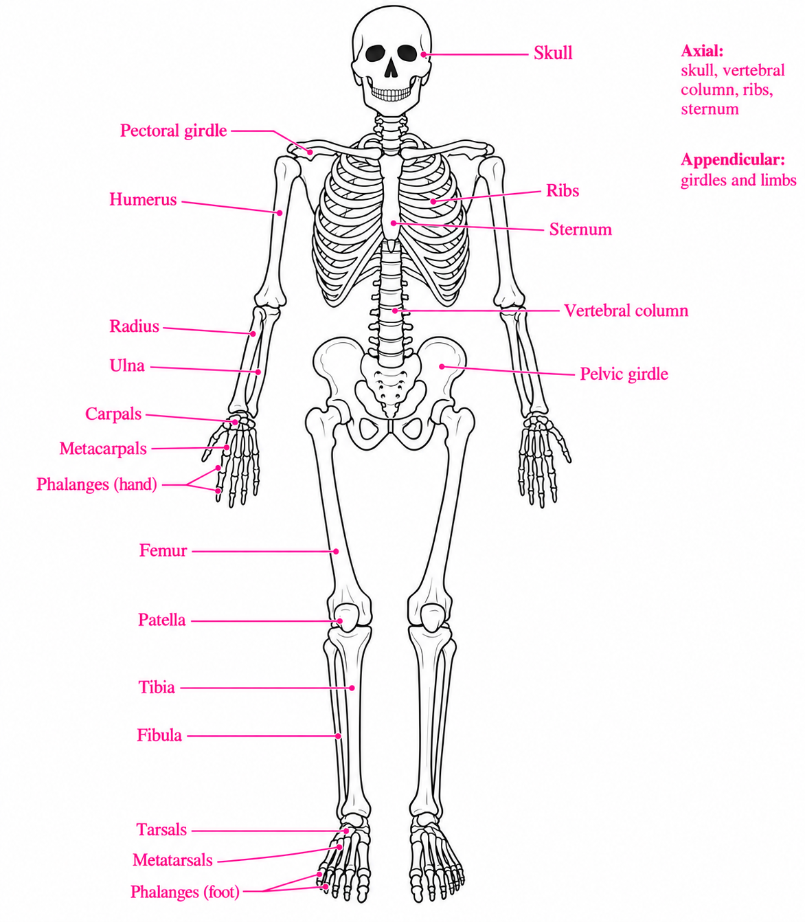
Human axial and appendicular skeleton with correctly labeled girdles, hand bones, and foot bones
  • Axial skeleton

– skull, vertebral column, sternum, ribs

  • Appendicular skeleton

– girdles: pectoral (clavicles and scapulae) and pelvic (hip bones)
– arm bones: humerus, radius, ulna

– hand bones: carpals, metacarpals, phalanges

– leg bones: femur, patella, tibia, fibula

– foot bones: tarsals, metatarsals, phalanges

3. Structure of bone

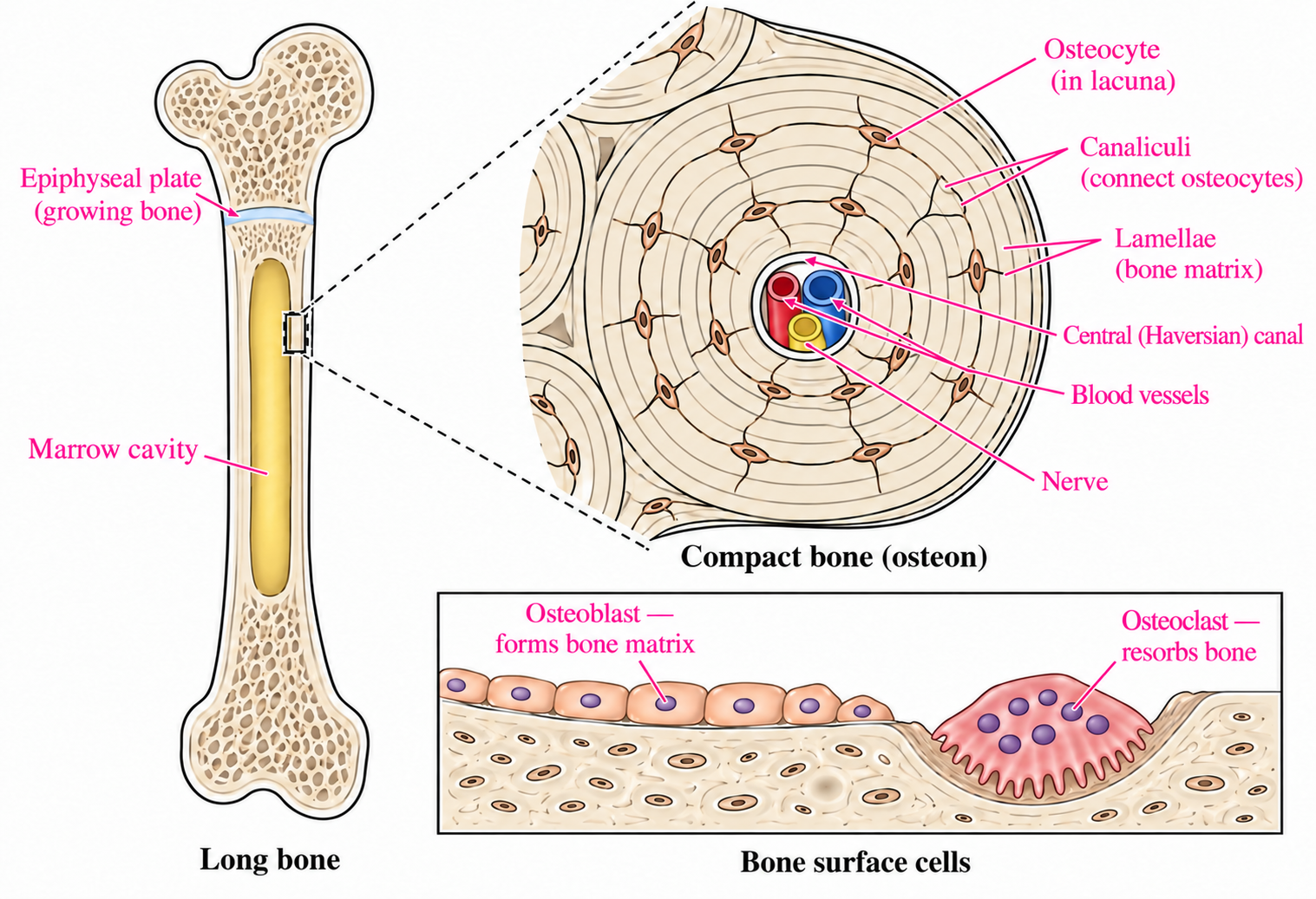
Growing long bone, compact bone osteon, and distinct bone-forming osteoblasts and bone-resorbing osteoclasts

Classification

Functions and features

Osteoclast

• Resorbs bone by dissolving mineral with acid and degrading the organic matrix; this can release calcium into the blood

Osteoblast

• Forms bone by secreting osteoid (organic matrix) and promoting its mineralization

Osteocyte

• A mature bone cell derived from an osteoblast; maintains bone matrix and senses mechanical stress

Haversian canal

• The central canal of an osteon; contains blood vessels and nerves. Canaliculi connect osteocytes with one another and with the central canal.

Epiphyseal plates

• Hyaline cartilage growth plates between the epiphyses and diaphysis of growing long bones; allow growth in length and close at skeletal maturity

E. Ear

1. Structure of ear

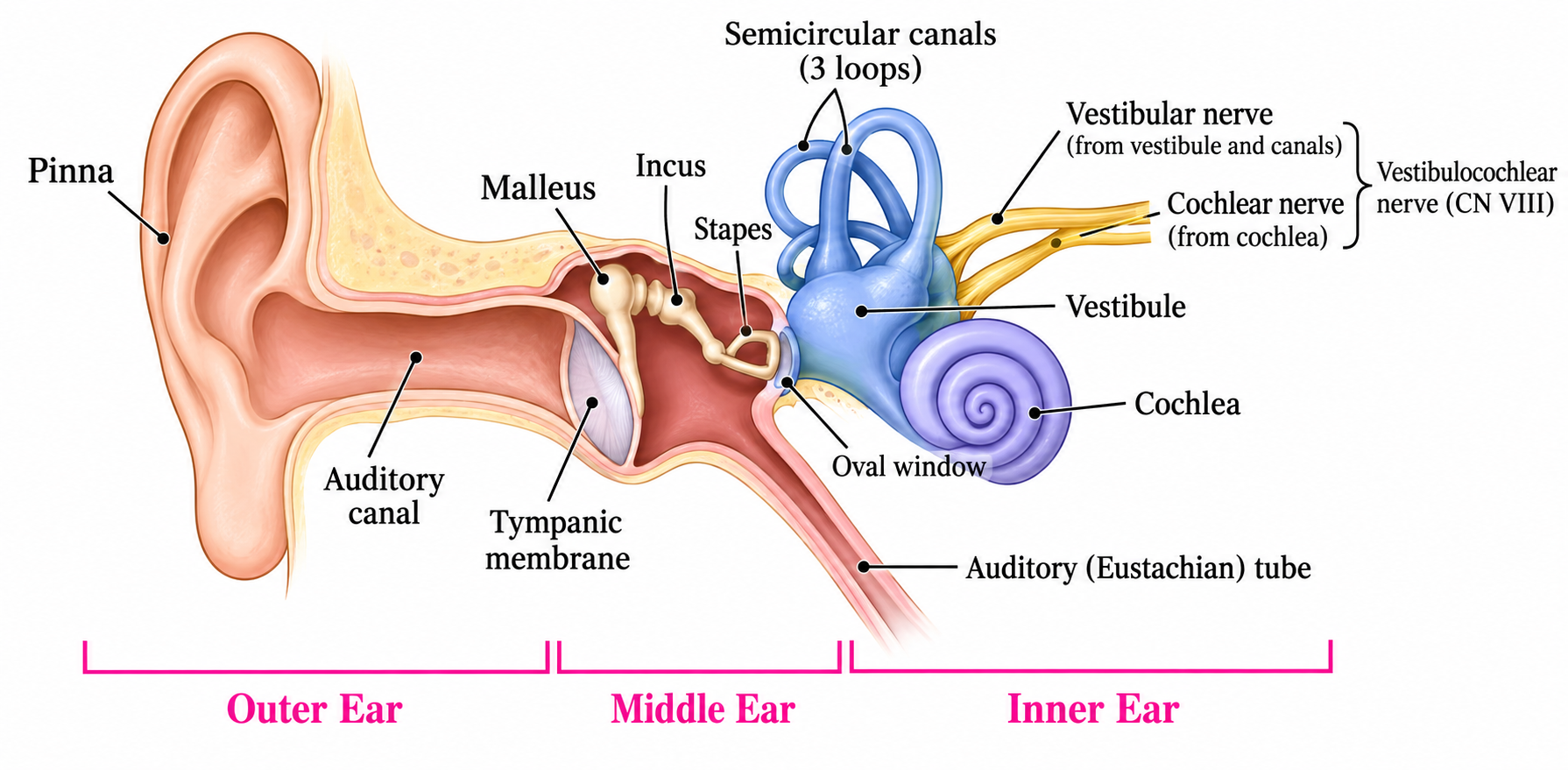
Outer, middle, and inner ear with ossicles, cochlea, vestibule, semicircular canals, and vestibulocochlear nerve

a. Outer ear

  • Collects outside sounds
  • Consists of pinna and auditory canal

b. Middle ear (ossicles)

  • Transmits vibrations from the eardrum to the oval window and amplifies pressure at the oval window
  • Consists of three small bones (ossicles): malleus, incus, and stapes

c. Inner ear

  • Fluid-filled chambers
  • Consists of the cochlea, vestibule (utricle and saccule), and semicircular canals. The cochlea detects sound; the utricle and saccule detect linear acceleration and head position, and the semicircular canals detect rotational acceleration.

Classification

Functions and features

Outer

 ear

Pinna

• Collects sound waves

Auditory canal

• Transmission of sound to tympanic membrane

Tympanic membrane

• Eardrum

• Separates the outer ear from the middle ear

• Vibrates in response to sound and transmits vibrations to the malleus

Middle

 ear

(ossicles)

Malleus

• Hammer shaped small bone

Incus

• Anvil shaped small bone

Stapes

• Stirrup shaped small bone

Inner

 ear

Semicircular

canals

• Three loop-shaped canals

• Detect rotational acceleration of the head and contribute to balance

Cochlea

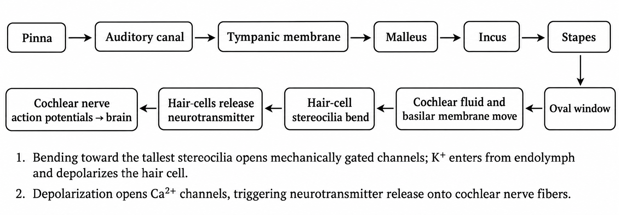
• Basilar membrane vibrations deflect hair-cell stereocilia; hair cells convert this mechanical stimulus into receptor potentials and release neurotransmitter onto cochlear nerve fibers

• Different regions respond best to different sound frequencies

Auditory nerve

• Transmission of sound signals to brain

2. Processing sound information

Sound transmission from pinna through ossicles to cochlea and hair-cell transduction into cochlear nerve signals

F. Eye

1. Structure of eye

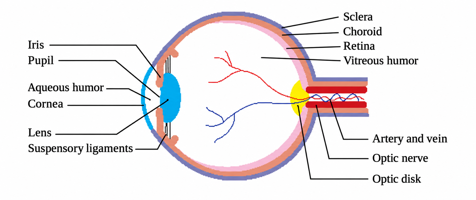
Eye cross-section with cornea, lens, suspensory ligaments, retinal layers, and optic nerve

Classification

Functions and features

Sclera

• The tough, white outer coat of the eye; protects the eye and helps maintain its shape

Cornea

• The transparent anterior part of the outer fibrous coat, continuous with the sclera

• Transmits light and provides most of the eye’s refractive power

Choroid

• A pigmented, vascular layer between the sclera and retina; nourishes the outer retina and absorbs stray light

Retina

• The innermost neural layer; contains photoreceptors and neurons that begin processing visual information

Iris

• Controls the amount of light by regulating pupil size

Pupil

• The central opening in the iris through which light enters the eye

Lens

• Focuses light on the retina

Aqueous humor

• Watery fluid in the anterior cavity between the cornea and lens; nourishes the cornea and lens and helps maintain intraocular pressure

Vitreous humor

• A transparent gel in the posterior cavity behind the lens; helps maintain eye shape and supports the retina

Optic disk

• The optic nerve exit site; lacks rods and cones and forms the blind spot

2. Light response in retina

a. Rod and cone cells

  • Rod cells – provide grayscale vision in dim light; their photopigment is rhodopsin
  • Cone cells – provide color vision and high visual acuity in bright light; their photopigments are cone opsins (photopsins)    

b. Light and dark responses

  • Response in light

– light activates photopigment (rhodopsin in rods; cone opsins in cones), which activates transducin and phosphodiesterase, lowering cGMP

– cGMP-gated cation channels close, reducing Na+ and Ca2+ influx

– rod and cone cells are hyperpolarized

– reduced glutamate release

  • Response in dark

– photopigment is inactive, and cGMP levels remain relatively high

– cGMP-gated cation channels are open, allowing Na+ and Ca2+ influx

– rod and cone cells are depolarized

– glutamate is released continuously