
VIII. Animal Physiology
Key focus of this chapter: digestive hormones
This chapter focuses on digestive hormones and gives concise summaries of key concepts in respiration, the cardiovascular system, osmoregulation and excretion, endocrinology, and nutrients.
A. Respiration
: process of gas exchange in the body.
- Pulmonary ventilation
– movement of air into and out of the lungs.
- External respiration
– exchange of O2 and CO2 between the alveoli and pulmonary capillary blood.
- Gas transport
– transport of O2 and CO2 in the blood between the lungs and body tissues.
- Internal respiration
– exchange of O2 and CO2 between systemic capillary blood and body tissues.
- Cellular respiration
– metabolic reactions that produce ATP; aerobic respiration uses oxygen and includes mitochondrial processes.
- Figure of external respiration

1. Respiratory tract
a. Air Passage


b. Function of each part
Classification | Features and roles |
Pharynx | • Muscular passageway for food and air |
Larynx | • Cartilaginous organ that maintains an open airway • Contains the vocal folds for sound production • Connects the pharynx to the trachea, anterior to the esophagus |
Epiglottis | • Flap of elastic cartilage that helps keep food out of the airway during swallowing |
Trachea | • Supported by C-shaped cartilage rings that keep the airway open • Windpipe |
Bronchi | • Conduct air into each lung |
Bronchioles | • Smaller branches of the bronchi • Fine airway tubes |
Alveoli | • Thin-walled air sacs where gases diffuse between air and pulmonary capillary blood |
2. Inspiration and expiration
a. Inspiration
- The diaphragm contracts and moves downward
- Thoracic volume increases, lowering alveolar pressure below atmospheric pressure
- Air flows into the alveoli down this pressure gradient
b. Expiration
- During quiet expiration, the diaphragm relaxes and moves upward
- Elastic recoil decreases thoracic volume, raising alveolar pressure above atmospheric pressure
- Air flows out of the alveoli down this pressure gradient
B. Cardiovascular System
1. Animal circulatory systems
Animal | Number of heart chambers | Number of Circuits |
Fish | Two | One |
Amphibians and most noncrocodilian reptiles (lizards, turtles, snakes) | Three | Two |
Birds, mammals, and crocodilians | Four | Two |
2. Mammalian heart
a. The pathway of circulation


b. Anatomy of the heart
Classification | Features and roles | |
Atrioventricular valves | • Allow blood to flow from an atrium to a ventricle • Prevent backflow into atrium • Flap composed of connective tissue | |
Semilunar valves | • Allow blood to flow from a ventricle into the aorta or pulmonary trunk • Prevent backflow into ventricle • Flap composed of connective tissue | |
Intercalated disks | • Junctional complexes between cardiac muscle cells, including gap junctions, desmosomes, and adherens junctions • Gap junctions allow ion flow that coordinates electrical activation; mechanical junctions hold cells together during contraction | |
Atrium | • Chamber for receiving blood | |
Ventricle | • Chamber for pumping blood out | |
Conduction system | Sino-atrial node (SA node) | • Functions as pacemaker • Located in the wall of the right atrium • Initiates impulses that spread through the atria and cardiac conduction system to coordinate heart contraction |
Atrioventricular node (AV node) | • Located in the inferior right atrium near the interatrial septum and tricuspid valve • Delays conduction, allowing atrial contraction to finish before ventricular contraction | |
c. Cardiac pressure
- Systole – phase of ventricular contraction
- Diastole – phase of ventricular relaxation
- Cardiac output
– volume of blood pumped by one ventricle per minute
– stroke volume × heart rate
- Stroke volume
– volume of blood pumped by one ventricle per beat
3. Blood vessels
a. Blood circulation in the blood vessels

b. Functions of each part
Blood vessels | Features and functions |
Artery | • Thick muscular wall and large vessel • Transports blood from heart to arteriole • Highest pressure |
Arteriole | • Small vessel • Transports blood from artery to capillary • Branched from artery • Determines the distribution of blood |
Capillary | • The smallest vessels, with a single endothelial cell layer supported by a basement membrane • Greatest total cross-sectional area and lowest average blood velocity across the capillary network • Connect arterioles to venules in tissue capillary beds • Exchange gases, nutrients, and wastes with surrounding tissues |
Venule | • Thin and small vessel • Collects blood from capillary • Skeletal muscle contractions help return venous blood toward the heart |
Vein | • Large vessel with low blood pressure • Transports blood from venules to heart • Many veins, especially in the limbs, contain valves that limit backflow |
** Portal venous system
- Carries blood from one capillary bed to another through portal veins
- Example: the hepatic portal vein
c. Blood pressure
- Blood flows down a pressure gradient, from higher to lower pressure; vascular resistance also affects flow.
d. Starling forces
- Driving forces for the fluid movement into and out of capillaries
- Capillary and interstitial hydrostatic pressures, together with plasma and interstitial oncotic pressures, influence fluid exchange.
Starling forces | Features and functions |
Capillary hydrostatic pressure | • Favors fluid movement from capillaries into interstitial fluid. • Hydrostatic pressure generally falls along a capillary; net fluid movement also depends on opposing pressures and capillary permeability. |
Capillary oncotic pressure (Osmotic pressure) | • Favors inward fluid movement and opposes filtration. • Plasma proteins, mainly albumin, generate capillary oncotic pressure. • In most tissues at steady state, lymphatics return filtered fluid; sustained reabsorption at the venous end is not universal. |

4. Blood
a. Cellular elements (45 % of blood)
Cell types | Features and functions | |
Erythrocytes (Red blood cells) | • Have hemoglobin to transport oxygen and some carbon dioxide • Mature mammalian red blood cells lack nuclei and mitochondria | |
Leukocytes (White blood cells) - cells of immune system - have nuclei - larger than red blood cells - can move into the lymphatic system from the blood vessels | Basophils | • Release histamine and heparin during allergic and inflammatory responses |
Eosinophils | • Minor phagocytic activity • Release granule contents that help kill parasites • Granule contents can also damage tissues during allergic inflammation | |
Lymphocytes | • Helper T cells – secrete cytokines that help activate B cells and cytotoxic T cells • B cells – form plasma cells that secrete antibodies • Cytotoxic T cells – use perforin and granzymes to trigger apoptosis in infected or tumor cells • Natural killer cells - use cytotoxic granules to kill infected or tumor cells | |
Monocytes | • Can differentiate into macrophages in tissues and carry out phagocytosis | |
Neutrophils | • Phagocytose microorganisms and foreign particles | |
Platelets | • Cell fragments derived from megakaryocytes that help stop bleeding (hemostasis) • Lack nuclei | |
b. Plasma elements (55 % of blood)
Classification | Features and functions | |
Ions | • Help regulate osmotic balance, membrane potentials, and pH • Examples: bicarbonate, chloride, sodium, potassium, calcium, and magnesium | |
Plasma proteins | Albumin | • Maintains plasma oncotic pressure, transports substances, and contributes to pH buffering |
Fibrinogen | • Converted to fibrin during blood clotting | |
Immunoglobulins | • Antibodies for immune response | |
Water | • Solvent for transporting other materials | |
Other substances | • Nutrients, hormones, waste products | |
c. Oxygen transport
- Hemoglobin
– is a tetrameric protein in red blood cells.
– an allosteric protein.
– each subunit has a heme group, which contains iron.
– each heme group binds O2 for transport.
– hemoglobin has a greater binding affinity for CO than for O2.
** Myoglobin
– binds O2 for storage in muscle.
– has a single globin subunit and stores oxygen in muscle.
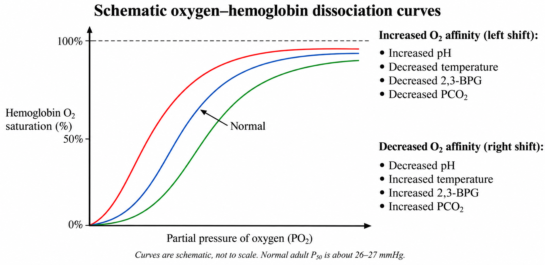
- Oxygen-binding curve of hemoglobin
– increased O2 binding affinity means increased percent O2 saturation at the same partial pressure of O2
– oxygen binding to hemoglobin is cooperative.
– the Bohr effect is a decrease in hemoglobin’s O2 affinity at lower pH, often associated with increased CO2.
d. Carbon dioxide transport
- Most CO2 (about 70%) from respiring cells is transported to lungs as bicarbonate ion (HCO3–) mainly in plasma. Most bicarbonate is formed in red blood cells by carbonic anhydrase and then moves into plasma in exchange for chloride
- About 23% of CO2 is bound to hemoglobin and transported
- The remaining CO2 (about 7%) is dissolved in plasma
- The bicarbonate buffer system helps maintain blood pH
- O2 and CO2 cross respiratory membranes by passive diffusion

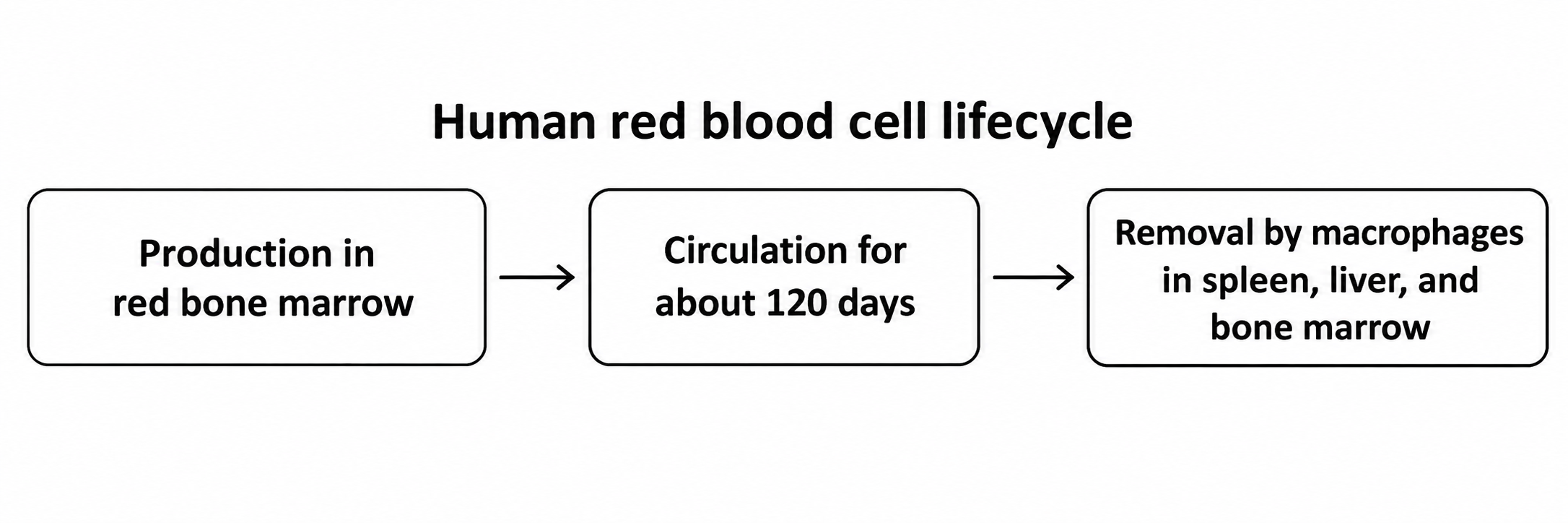
e. States of red blood cells
f. Blood clotting
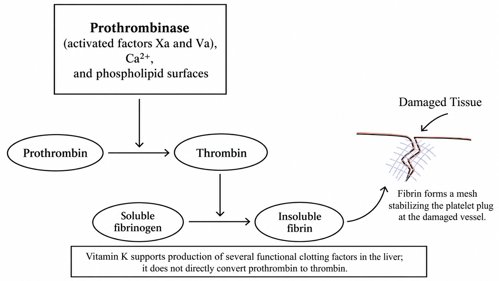
- ① A prothrombinase complex of activated clotting factors converts prothrombin to thrombin on phospholipid surfaces in the presence of Ca2+. Vitamin K is required for the liver to produce several functional clotting factors.
- ② Thrombin converts soluble fibrinogen into insoluble fibrin strands.
- ③ Fibrin forms a mesh that stabilizes the platelet plug at the damaged vessel.
C. Digestion
1. Mouth
- Mechanical breakdown of food by teeth.
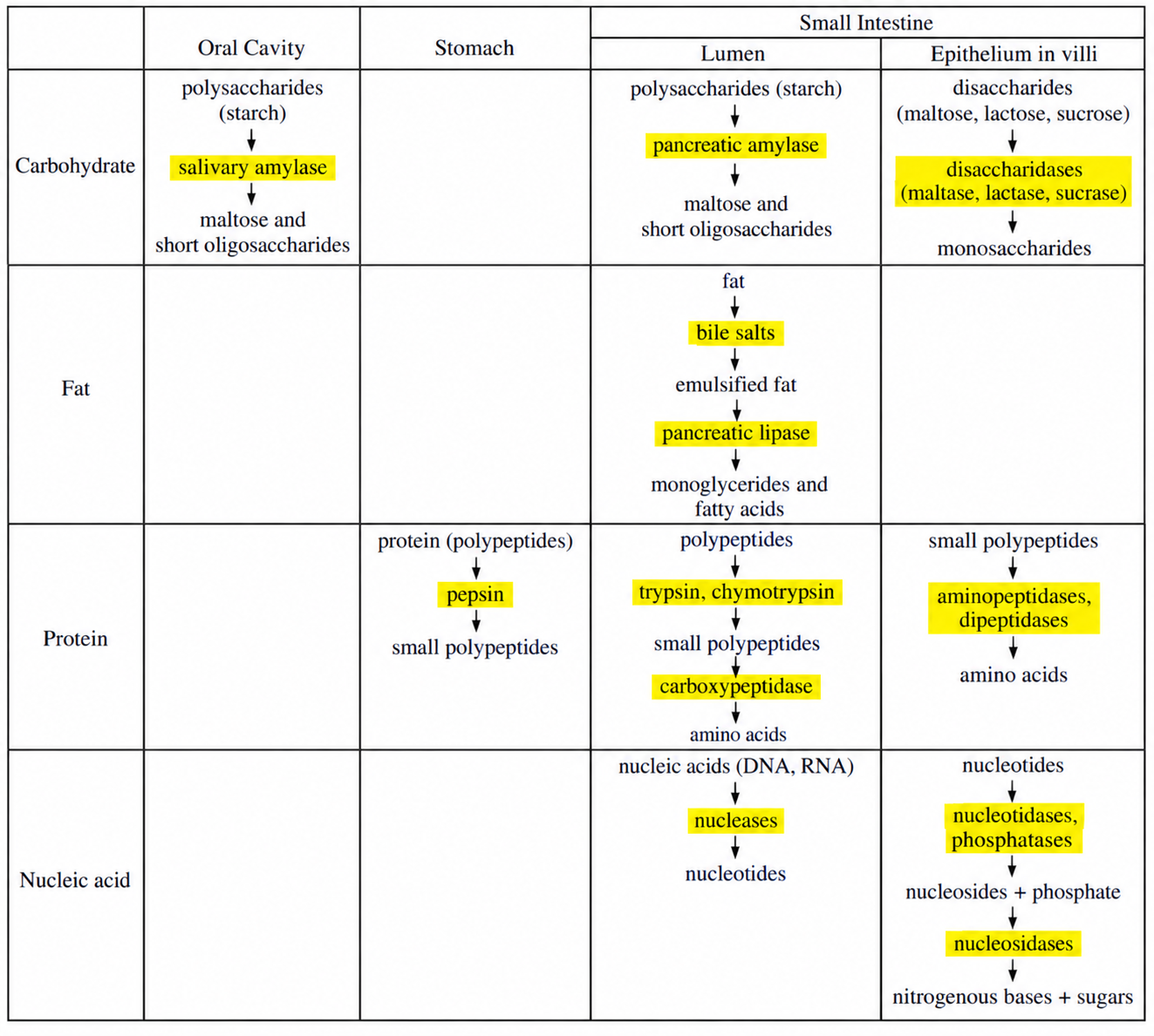
- Chemical breakdown by salivary amylase, which breaks down starch to disaccharides.
- Moistening of food to form a bolus.
2. Esophagus
- Conducts food from mouth to stomach by waves of muscular contraction called peristalsis.
- Controls the passage of food by muscular ring valves called sphincters.
3. Stomach
- Stores food and supports digestion.
- Mechanical breakdown by churning movements.
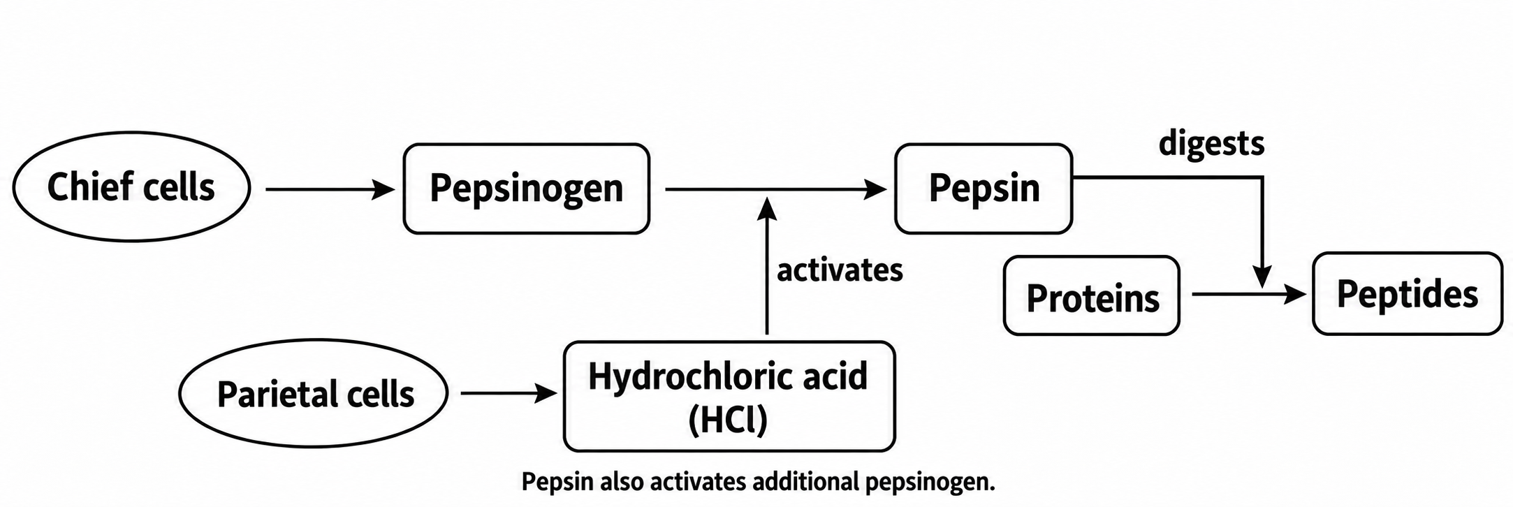
- Gastric glands secrete mucus from mucous cells, pepsinogen from chief cells, and hydrochloric acid (HCl) from parietal cells.
- Pepsin, which is converted from pepsinogen by HCl, digests proteins.
- Food mixed with gastric juice becomes acid chyme.
- Mucus lubricates and protects the surface of stomach walls.
- The lower esophageal (gastroesophageal) sphincter helps prevent backflow from the stomach into the esophagus.
- Pyloric sphincter controls the passage of acid chyme from stomach to small intestine.
4. Small intestine
: most chemical digestion occurs in the small intestine, which consists of the duodenum, jejunum, and ileum.
a. Secretion to duodenum
- Pancreas – secretes pancreatic juice, which contains the digestive enzymes and bicarbonate for neutralizing the acid chyme, to duodenum.
- Liver – produces bile; bile salts emulsify fats and aid their digestion and absorption in the small intestine.
- Small-intestinal epithelium – produces brush-border digestive enzymes.
b. Absorption
- Villi – mucosal projections that increase the surface area for absorption.
- Microvilli – microscopic projections on the surfaces of individual epithelial cells.
- Long-chain fatty acids and monoglycerides enter intestinal cells, are reassembled into triglycerides, and are packaged into chylomicrons that enter lacteals and reach the blood through lymph. Glycerol and short-chain fatty acids enter portal blood directly.
- Glucose and amino acids enter blood capillaries and travel to the liver through the hepatic portal circulation.

5. Liver
a. Secretion for digestion
- Produces bile, which is stored in the gallbladder.
- Bile salts emulsify fats and support lipid digestion; bile does not contain digestive enzymes.
b. Multiple functions
- Destruction of aged red blood cells.
- Glycogen synthesis, breakdown, and storage.
- Synthesis of plasma proteins such as albumin, clotting proteins, and angiotensinogen.
- Conversion of ammonia into urea for excretion (detoxification).
- Removal of wastes and bacteria from the blood.
6. Pancreas
a. Exocrine (secretion of pancreatic juice into the duodenum)
- Secretes amylase to digest carbohydrates.
- Secretes trypsinogen, which enteropeptidase activates to trypsin in the small intestine for protein digestion.
- Secretes lipase to digest fats.
- Secretes bicarbonate ions to neutralize the acid chyme.
b. Endocrine (secretion of hormones into the bloodstream)
- α cells secrete glucagon, which stimulates hepatic glycogen breakdown and gluconeogenesis.
- β cells secrete insulin, which promotes glucose uptake in muscle and adipose tissue and glycogen storage in liver and muscle.
7. Large intestine
- The large intestine includes the cecum, colon, rectum, and anal canal. The colon has ascending, transverse, descending, and sigmoid regions.
- Absorption of water, ions, and other minerals.
- Feces transformation and storage of feces.
- Gut bacteria produce vitamin K; dietary vitamin K is absorbed mainly in the small intestine.
8. Overview of digestion
9. Hormonal control of digestion

D. Osmoregulation and excretion
- Osmoregulation – regulates the water and solute balance in body fluids.
- Excretion – removes metabolic wastes, including nitrogen-containing wastes, from the body.
1. Animal osmoregulation and excretion
Animal group | Osmoregulation and excretion | |
Flatworms | • Protonephridia with flame cells | |
Earthworm | • Metanephridia | |
Insect | • Malpighian tubules | |
Bony fish | Freshwater fish | • Excrete large volumes of dilute urine and actively absorb ions through the gills to balance water gain in a hypotonic environment |
Saltwater fish | • Drink seawater to replace water lost by osmosis and excrete excess salts through the gills and urine | |
Bird | • Marine birds use nasal salt glands to excrete concentrated salt solutions, allowing them to drink seawater | |
2. Mammalian osmoregulation and excretion
a. Basic renal processes in the mammalian kidney

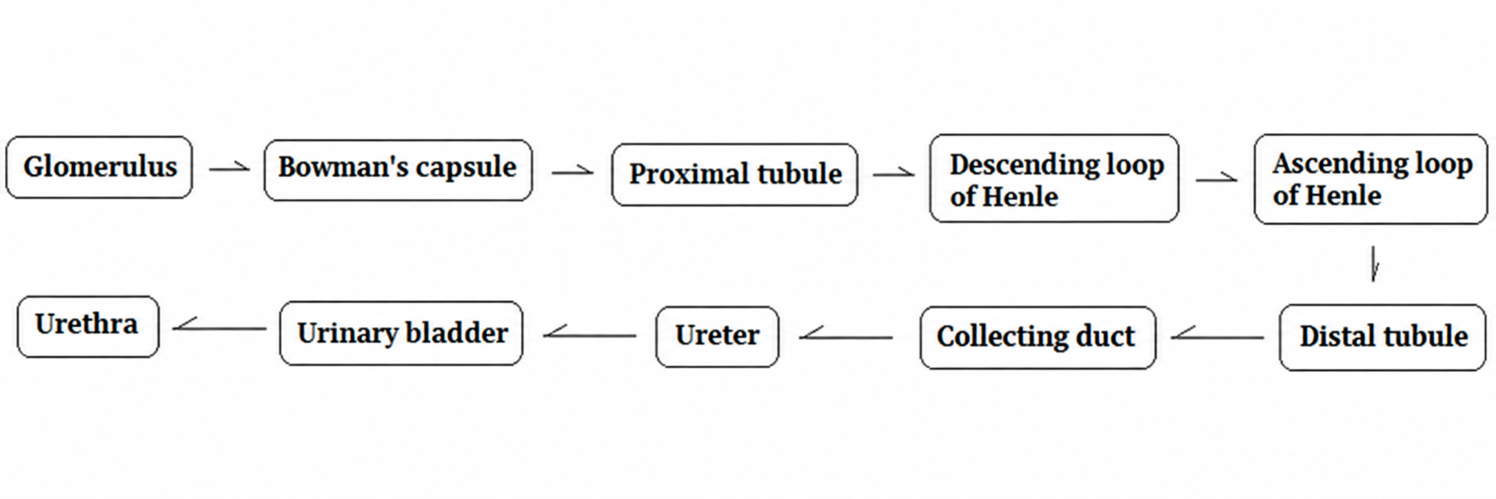
- Filtration – pressure-driven passage of water and small solutes from glomerular blood into Bowman’s capsule; blood cells and most plasma proteins remain in the blood.
- Secretion – moves substances from peritubular blood into the renal tubules by active or passive transport.
- Reabsorption – returns filtered substances from the renal tubules to the blood by active or passive transport.
- Excretion – eliminates substances in urine. The amount excreted equals the amount filtered minus the amount reabsorbed plus the amount secreted.
b. Function of each part
Kidney processes | Features and functions | |
Filtration (pressure-driven) | Glomerulus | • Tuft of capillaries • Glucose is freely filtered across the glomerular filtration barrier |
Bowman’s capsule | • Cup-shaped structure at the beginning of the nephron • Surrounds the glomerulus • Receives filtrate containing water, glucose, and small solutes such as ions, amino acids, and urea • The filtration barrier retains blood cells and most plasma proteins | |
Secretion (active or passive transport) and Reabsorption ( active or passive transport) | Proximal tubule | • Reabsorbs water, salt, and nutrients such as glucose and amino acids; secretes selected solutes • Reabsorbs most filtered water and salt • Contributes to pH regulation |
Distal tubule | • Adjusts salt reabsorption and secretion • Regulates potassium and sodium balance • Contributes to pH regulation • Aldosterone increases sodium reabsorption and potassium secretion in the late distal nephron; ADH regulates water permeability | |
Reabsorption transport) | Descending loop of Henle | • Reabsorption of water |
Ascending loop of Henle | • Reabsorbs NaCl and is impermeable to water | |
Excretion | Collecting duct | • Receives tubular fluid and helps determine final urine concentration through ADH-regulated water reabsorption and medullary urea recycling |

c. Passage of waste products
E. Endocrinology
1. Hormones
: chemical messengers released by endocrine cells into the bloodstream that act on target cells with the appropriate receptors.
a. Hypothalamic hormones
- Hypothalamic releasing and inhibiting hormones regulate the anterior pituitary through portal blood vessels. The hypothalamus also synthesizes ADH and oxytocin, which are released from the posterior pituitary.
b. Steroid hormones
- Lipid-soluble and poorly soluble in water.
- Cross plasma membranes and bind to receptors in the cytoplasm or nucleus.
- Examples: androgens, estrogens, progesterone, cortisol, and aldosterone
c. Peptide hormones
- Hydrophilic and dissolved in water.
- Generally cannot diffuse through the lipid bilayer and act through receptors on the target cell surface.
- Examples: insulin, glucagon, and most pituitary hormones
d. Tropic hormones
- Act on other endocrine glands to regulate their hormone secretion.
- Examples: FSH, LH, TSH, and ACTH
e. Nontropic hormones
- Act primarily on nonendocrine target tissues; examples include endorphins, MSH, and prolactin.
2. Hormones of endocrine glands

Hypothalamic hormones | Pituitary | Hormone | Target | Functions of pituitary hormones |
GHRH (stimulates GH) GHIH (inhibits GH) | Anterior Pituitary | GH (growth hormone) | Liver, bone, and other tissues | • Stimulates growth through direct effects and by promoting IGF-1 production, mainly in the liver |
TRH (stimulates TSH) | TSH (thyroid-stimulating hormone) | Thyroid | • Stimulates thyroid growth • Stimulates production and release of T3 and T4 • T3 and T4, rather than TSH directly, regulate cellular metabolism | |
CRH (stimulates ACTH) | ACTH | Adrenal cortex | • Participates in the hypothalamic-pituitary-adrenal stress response • Primarily stimulates glucocorticoid secretion, especially cortisol; aldosterone is regulated mainly by angiotensin II and potassium | |
GnRH (stimulates LH and FSH) | LH | Testis and ovary | • Triggers ovulation and corpus luteum formation • Stimulates Leydig cells to secrete testosterone in the testes | |
FSH (follicle-stimulating hormone) | • Stimulates growth of antral ovarian follicles and estrogen production by granulosa cells • Acts on Sertoli cells to support spermatogenesis in the testes | |||
PRH (stimulation) PIH (dopamine) (inhibition) | Prolactin | Mammary glands | • Stimulates milk production by the mammary glands | |
MSH | Melanocytes | • Stimulates color changes of skin in reptiles and amphibians | ||
Posterior Pituitary | ADH (antidiuretic hormone, vasopressin) | Kidney tubules | • Increases water reabsorption in the late distal nephron and collecting ducts • Produces more concentrated urine and helps maintain blood volume and pressure • Insufficient ADH causes central diabetes insipidus | |
Oxytocin | Mammary glands, uterine muscles | • Initiates milk release • Induces labor |
3. T3 and T4 from the thyroid gland
: the thyroid gland has two lobes on either side of the trachea. Its hormones regulate cellular metabolism and influence functions such as digestion, heart rate, blood pressure, and reproduction.
- Thyroid follicular cells secrete T3 and T4 under stimulation by pituitary TSH.

- Triiodothyronine (T3)
– has three iodine atoms.
– has greater biological activity than T4.
- Thyroxine (T4)
– has four iodine atoms.
– is produced in greater amounts than T3.
– converted to T3 in the kidneys, liver, and other target tissues.
- Metabolic disorders of the thyroid gland
Metabolic disorders | Causes and symptoms |
Hyperthyroidism | • Excessive thyroid hormone production • Increased metabolic rate with heat intolerance, irritability, sweating, rapid heartbeat, and weight loss • Example: Graves’ disease, which can cause protruding eyes |
Hypothyroidism | • Insufficient thyroid hormone production • Decreased metabolic rate with cold intolerance, lethargy, and weight gain • Severe hypothyroidism may cause myxedema; hypothyroidism present at birth is called congenital hypothyroidism |
4. Regulation of blood calcium levels
a. Parathyroid hormone (PTH)
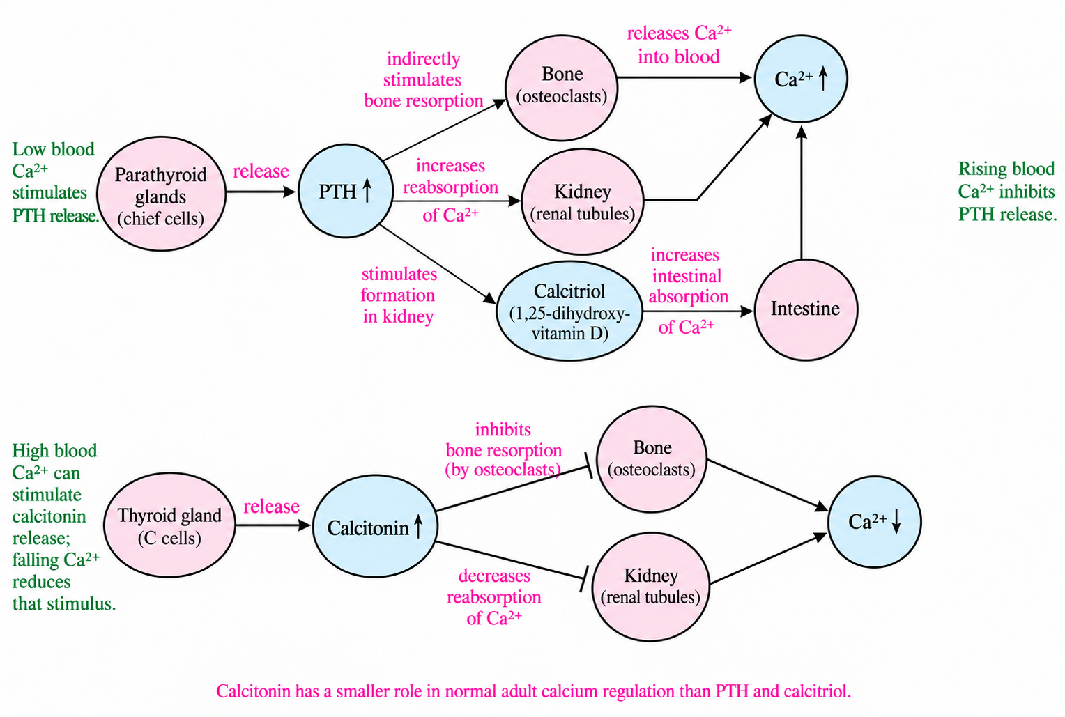
- Secreted by the parathyroid glands when Ca2+ levels in the blood fall.
- Indirectly stimulates osteoclast-mediated bone resorption, releasing calcium into the blood.
- Increases renal calcium reabsorption and stimulates formation of calcitriol, the active form of vitamin D, which increases intestinal calcium absorption.
b. Calcitonin
- Secreted by thyroid parafollicular (C) cells when Ca2+ levels in the blood rise.
- Inhibits osteoclast-mediated bone resorption, helping lower blood calcium.
- Can increase renal calcium excretion; its role in normal adult calcium regulation is smaller than that of PTH and calcitriol.
5. Regulation of blood glucose levels
a. Glucagon
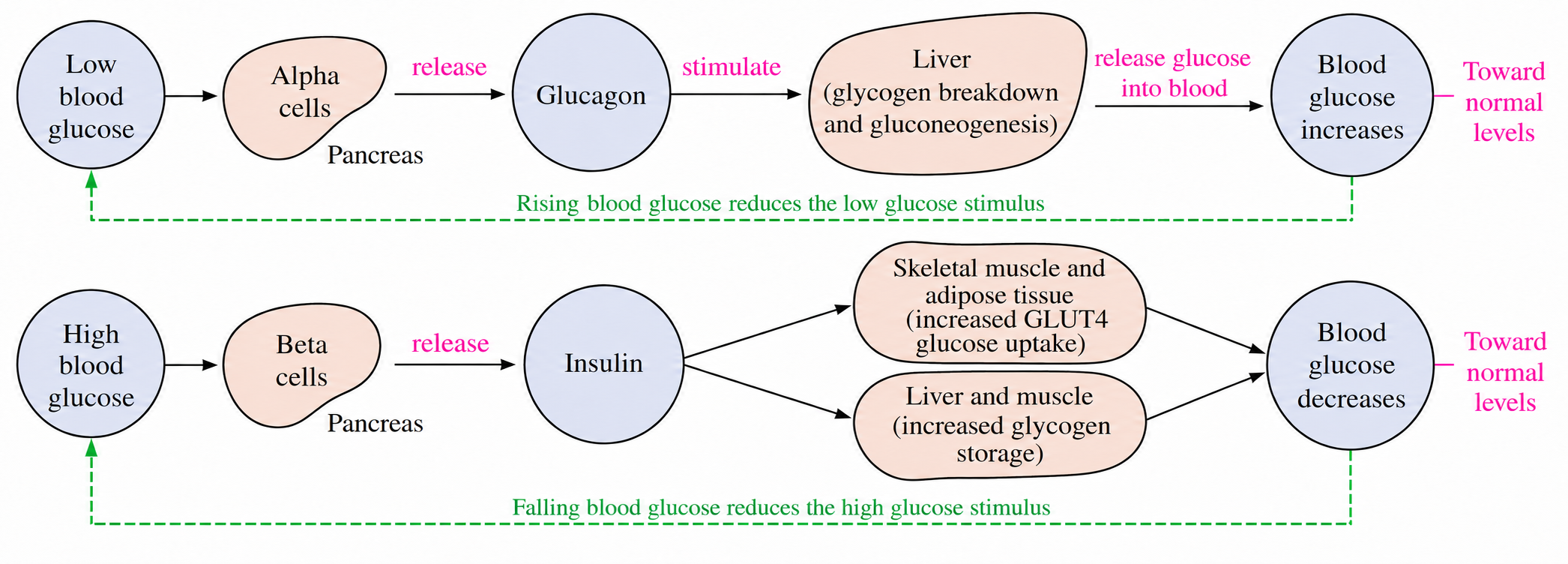
- Secreted by alpha cells in pancreas when glucose levels in blood are decreased.
- Stimulates hepatic glycogen breakdown and gluconeogenesis.
- Stimulates liver to release glucose into blood stream.
b. Insulin
- Secreted by beta cells in pancreas when glucose levels in blood are increased.
- Lowers blood glucose by increasing GLUT4-mediated glucose uptake in skeletal muscle and adipose tissue and promoting glucose storage.
- Promotes glycogen synthesis in liver and skeletal muscle and suppresses hepatic glucose production.
** Pancreatic islets (islets of Langerhans) contain alpha cells, beta cells, delta cells, and pancreatic polypeptide (F) cells.
6. Stress response
- Regulated in part by the adrenal glands, located above the kidneys
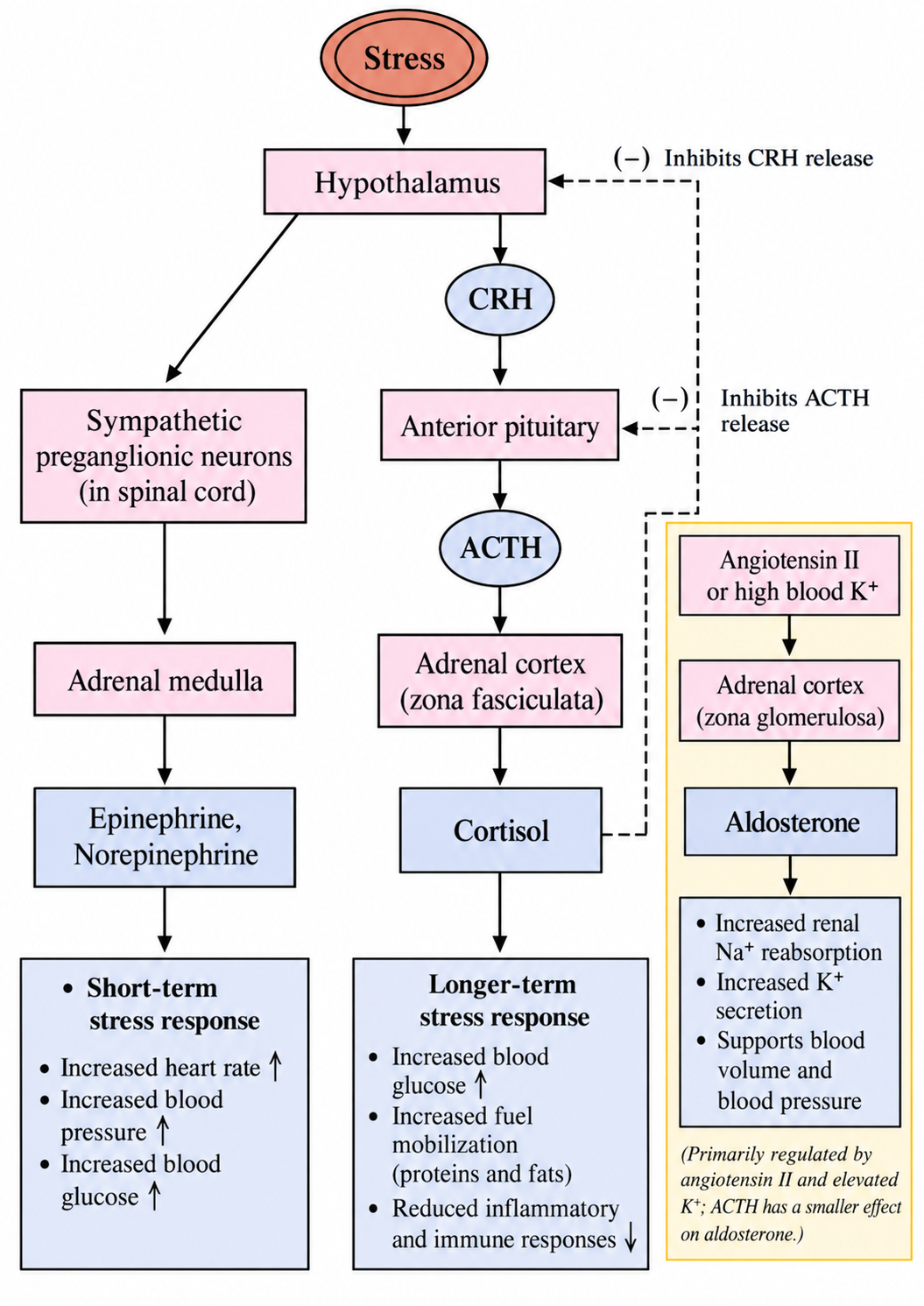
a. Adrenal medulla
- Receives sympathetic stimulation through spinal cord pathways under hypothalamic control.
- Secretes epinephrine and norepinephrine in response to short-term stress.
- These hormones raise blood glucose, heart rate, blood pressure, and metabolic activity.
b. Adrenal cortex
- Cortisol secretion is stimulated by ACTH.
- Aldosterone secretion is regulated mainly by angiotensin II and elevated blood potassium; ACTH has a smaller effect.
- Cortisol increases blood glucose availability and suppresses inflammatory and immune responses.
- Aldosterone increases renal sodium reabsorption and potassium secretion, helping maintain blood volume and pressure.
- Water can follow retained sodium by osmosis; ADH regulates water permeability in the collecting ducts.
7. Other chemical messengers
a. Pheromones
- Chemical signals released outside the body that affect other members of the same species.
- Can function in territorial marking, courtship, and social organization within a species.
b. Renin
- Proteolytic enzyme that regulates arterial blood pressure.
- Often described as a hormone.
c. Melatonin
- A tryptophan-derived amine hormone secreted by the pineal gland in the brain.
- Functions in circadian rhythms.
- Secreted at night.
d. Endorphin
- A neuropeptide that acts as a chemical messenger.
- Decreases pain perception in the brain.
F. Nutrients
1. Vitamins
- Organic compounds
- Needed in small amounts; deficiencies can cause health problems
- Many vitamins must be obtained from the diet; humans can synthesize vitamin D and produce some niacin from tryptophan.
Vitamins | Types | Functions |
Water-soluble Vitamins | Vitamin B1 (thiamine) | • Forms thiamine pyrophosphate, a coenzyme used in carbohydrate metabolism, including oxidative decarboxylation • Contained in grains, peanuts, pork, and legumes |
Vitamin B2 (riboflavin) | • Component of coenzymes FAD and FMN • Supports energy metabolism • Contained in grains, vegetables, and meats | |
Vitamin B3 (niacin) | • Component of the coenzymes NAD+ and NADP+ • Contained in grains, nuts, and meats • Deficiency causes pellagra | |
Vitamin C (L-ascorbic acid) | • Involved in collagen synthesis • Supports skin and other connective tissues • Deficiency causes scurvy • Contained in fruits and vegetables | |
Fat-soluble Vitamins | Vitamin A (retinol) | • Retinal, derived from vitamin A, is the light-absorbing component of visual pigments • Deficiency can cause night blindness • Found as preformed vitamin A in animal foods and as provitamin A carotenoids in green and orange vegetables |
Vitamin D | • Promotes intestinal calcium and phosphate absorption and supports bone health and immune function • Produced in the skin following UVB exposure • Contained in egg yolk • Deficiency can cause rickets in children and osteomalacia in adults | |
Vitamin E (tocopherol) | • Acts as an antioxidant that protects cell membranes from oxidative damage • Deficiency can cause neurologic problems and hemolytic anemia • Contained in nuts, seeds, and vegetable oils | |
Vitamin K (phylloquinone) | • Cofactor needed to produce functional blood-clotting proteins • Dietary vitamin K is absorbed in the small intestine • Contained in green vegetables |
2. Minerals
- Inorganic substances
- Needed in appropriate amounts; both deficiency and excess can cause health problems
- Phosphorus, magnesium, calcium – important in bone and tooth formation
- Sodium and potassium – function in muscle contraction and nerve conduction
- Iron – helps bind oxygen in hemoglobin and is a component of electron carriers