VI. Embryology

Key focus of this chapter: male and female reproductive systems

This chapter reviews the male and female reproductive systems, fertilization, embryonic development, extraembryonic membranes in shelled eggs, and the mammalian placenta.

A. Male reproductive system

1. Anatomy

Male reproductive system

Functions

Testis

Seminiferous

Tubules

• Production of sperm

• Spermatogonia develop into sperm through spermatogenesis

Leydig cells (Interstitial cells)

• LH stimulates synthesis of androgens, primarily testosterone

Sertoli cell

• Nurturing and controlling sperm development

Epididymis

• Place for maturation and storage of sperm

Prostate gland

• Neutralizing the acidity of the vaginal tract and seminal fluid by secreting slightly alkaline milky fluid

• Enhancing motility and life of sperm

Seminal vesicle

• Secretes fructose-rich fluid that nourishes sperm and contributes most of the semen volume

Vas deferens

• Duct for expulsion of sperm

Ejaculatory duct

• Joining ducts of seminal vesicle and vas deferens

Urethra

• Duct of semen and urine

2. The passage of sperm

3. Sperm

  • Acrosome

– contains enzymes that help the sperm penetrate the oocyte’s surrounding layers during fertilization

  • Nucleus – contains the haploid paternal genome
  • Mitochondria – concentrated in the midpiece and supply ATP for motility
  • Flagellum

– movement of sperm

4. Hormone regulation

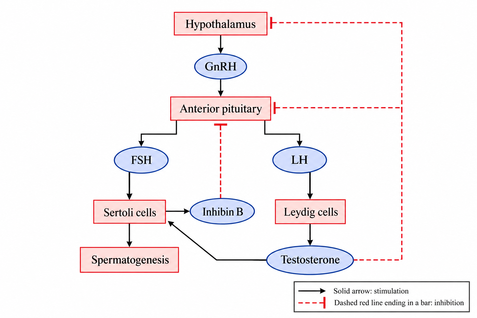
FSH acts on Sertoli cells to support spermatogenesis; LH acts on Leydig cells to stimulate testosterone production. Testosterone inhibits GnRH and LH secretion, while inhibin B from Sertoli cells inhibits FSH secretion.

B. Female reproductive system

1. Anatomy

Female reproductive system

Functions

Follicle

• An ovarian structure containing an oocyte surrounded by supporting follicular cells

Ovary

• Site of oogenesis and secretion of estrogen and progesterone

Ovulation

• A mature follicle ruptures and releases a secondary oocyte arrested in metaphase II

• The ruptured follicle becomes the corpus luteum

Corpus luteum

• Temporary endocrine structure formed from the ruptured follicle; secretes progesterone and estrogens

Oviduct (Fallopian tube)

• Place of fertilization or passage of egg

Uterus

• Site of blastocyst implantation in the endometrium

2. Passage of an oocyte

3. Hormone regulation

  • FSH

– supports sperm production in males and follicle growth in females.

– stimulates granulosa cells and the growth of antral follicles.

  • LH

– stimulates testes and ovaries.

– the LH surge triggers ovulation and formation of the corpus luteum.

  • Estrogen

– stimulates proliferation of the uterine lining before implantation of the blastocyst.

– promotes development of female secondary sex characteristics.

  • Progesterone

– maintains the uterine lining and promotes secretory changes that support implantation and pregnancy.

** Estrogen and progesterone usually provide negative feedback on FSH and LH secretion; sustained high estrogen before ovulation provides positive feedback that triggers the LH surge.

4. Comparison of oogenesis with spermatogenesis

a. Oogenesis

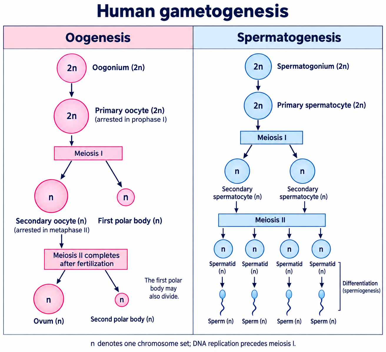
  • Each primary oocyte can produce one functional ovum and polar bodies; in humans, meiosis II is completed only after fertilization
  • Unequal distribution of the cytoplasm

b. Spermatogenesis

  • Each primary spermatocyte produces four haploid spermatids, which differentiate into spermatozoa
  • Cytoplasm equally distributed

C. Fertilization

1. External fertilization of sea urchin

a. Process of fertilization

  • ① Contact – The sperm head contacts the egg’s jelly coat.
  • ② Acrosomal reaction – Acrosomal enzymes digest a path through the jelly coat; the acrosomal process binds receptors on the vitelline layer.
  • ③ Depolarization – Fusion of the sperm and egg plasma membranes triggers rapid egg membrane depolarization, producing the fast block to polyspermy.
  • ④ Insertion of sperm nucleus into egg.
  • ⑤ Cortical reaction – A rise in egg cytosolic Ca2+ triggers cortical granule exocytosis, which modifies and elevates the vitelline layer into the fertilization envelope and causes a slow block to polyspermy.

b. Function of each part

Parts

Functions

Acrosome

• Contains hydrolytic enzymes that digest a path through the sea urchin egg’s jelly coat

Jelly coat

• Gelatinous outer egg covering

• Contact with the jelly coat triggers the sperm’s acrosomal reaction

Vitelline layer

• Extracellular matrix outside the egg plasma membrane and beneath the jelly coat

• Contains sperm-binding receptors

Sperm binding receptor

• Recognizes sperm bindin on the acrosomal process and mediates attachment to the vitelline layer

Cortical granules

• Release their contents by Ca²⁺-triggered exocytosis, modifying the vitelline layer to block polyspermy

2. Internal fertilization of mammals

a. Process of fertilization

  • ① Approaching – capacitated sperm pass through the surrounding follicle cells and approach the zona pellucida of the secondary oocyte.
  • ② Acrosomal reaction – acrosomal enzymes and sperm motility help the sperm penetrate the zona pellucida and reach the oocyte plasma membrane.
  • ③ The sperm and oocyte plasma membranes fuse.
  • ④ The sperm nucleus enters the oocyte; the oocyte completes meiosis II.
  • ⑤ Cortical reaction – a rise in oocyte cytosolic Ca2+ triggers cortical granule exocytosis, which modifies the zona pellucida and causes a slow block to polyspermy.

b. Function of each part

Parts

Functions

Acrosome

• Contains enzymes that help sperm penetrate the zona pellucida

Zona pellucida

• Glycoprotein extracellular coat surrounding the oocyte

• Participates in sperm recognition and binding

Cortical granules

• Release enzymes that modify the zona pellucida after sperm–oocyte fusion, helping prevent polyspermy

c. Twins

  • Identical (monozygotic) twins – one fertilized egg develops into two embryos
  • Fraternal (dizygotic) twins – two eggs are fertilized by two different sperm and develop into two embryos

d. Cell differentiation

  • Cells become more specialized in structure and function
  • A single zygote develops into a multicellular organism with specialized tissues

D. Embryonic development

1. Description of each stage

Stages

Description

Cleavage
(Zygote cell division)

Morula

• Rapid mitotic cleavages form a solid ball of cells without increasing overall embryo size

Blastula

• A blastula contains a fluid-filled cavity called the blastocoel

• In mammals, the blastocyst has a trophoblast and an inner cell mass

Gastrulation

• In amphibians, gastrulation begins at the dorsal lip of the blastopore

• Forms three germ layers (ectoderm, mesoderm, endoderm) and a primitive gut in triploblastic animals

• Gastrulation movements vary among animal groups

• Archenteron – the primitive gut cavity

• Blastopore – generally gives rise to the mouth in protostomes and the anus in deuterostomes

• Involution – surface cells roll inward over the blastopore lip in amphibians

Organogenesis

• Organ primordia develop from the three germ layers

• Neurulation – the neural plate folds to form a hollow neural tube, which develops into the brain and spinal cord

• Induction – signals from one tissue influence the development of another; the notochord helps pattern the overlying neural tissue

• Neural crest cells, somites and the notochord contribute to distinct developing structures

2. Three germ layers

Ectoderm

Mesoderm

Endoderm

• Epidermis of skin

• Sensory receptor of epidermis

• Nervous system

• Lens of eyes

• Teeth: enamel from oral ectoderm; other dental tissues from neural crest

• Adrenal medulla

• Circulatory system

• Kidneys and ureters

• Gonadal supporting tissues and most reproductive ducts (germ cells have a separate lineage)

• Lymphatic system

• Most connective tissue

 (Dermis of skin)

• Most skeletal tissue

• Muscular system

• Adrenal cortex

• Epithelial lining and glands of the lower respiratory tract

• Most digestive tract epithelium and associated glandular tissues, including liver and pancreas

• Most urinary bladder epithelium

• Muscle and connective tissue in these organs arise mainly from mesoderm

** Totipotent: a single cell can generate all embryonic and extraembryonic cell types needed to form a complete organism

E. Functions of extraembryonic membranes in the shelled eggs

Classification

Functions and features

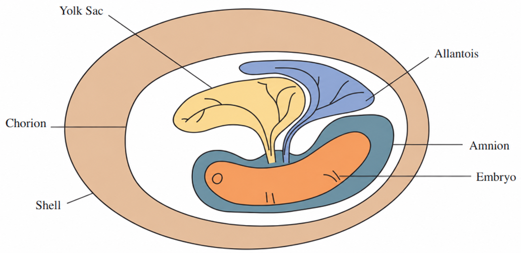
Amnion

• Membranous sac for protecting an embryo from mechanical shock and dehydration

• Not present in amphibians

Chorion

• With the allantois, supports oxygen and carbon dioxide exchange between the embryo and the external environment

Allantois

• Stores nitrogenous wastes in shelled amniotic eggs and supports gas exchange with the chorion

• In humans, contributes to umbilical structures and the urachus; the chorion forms the fetal portion of the placenta

Yolk sac

• Provides nutrients to embryo

F. Placenta in mammals

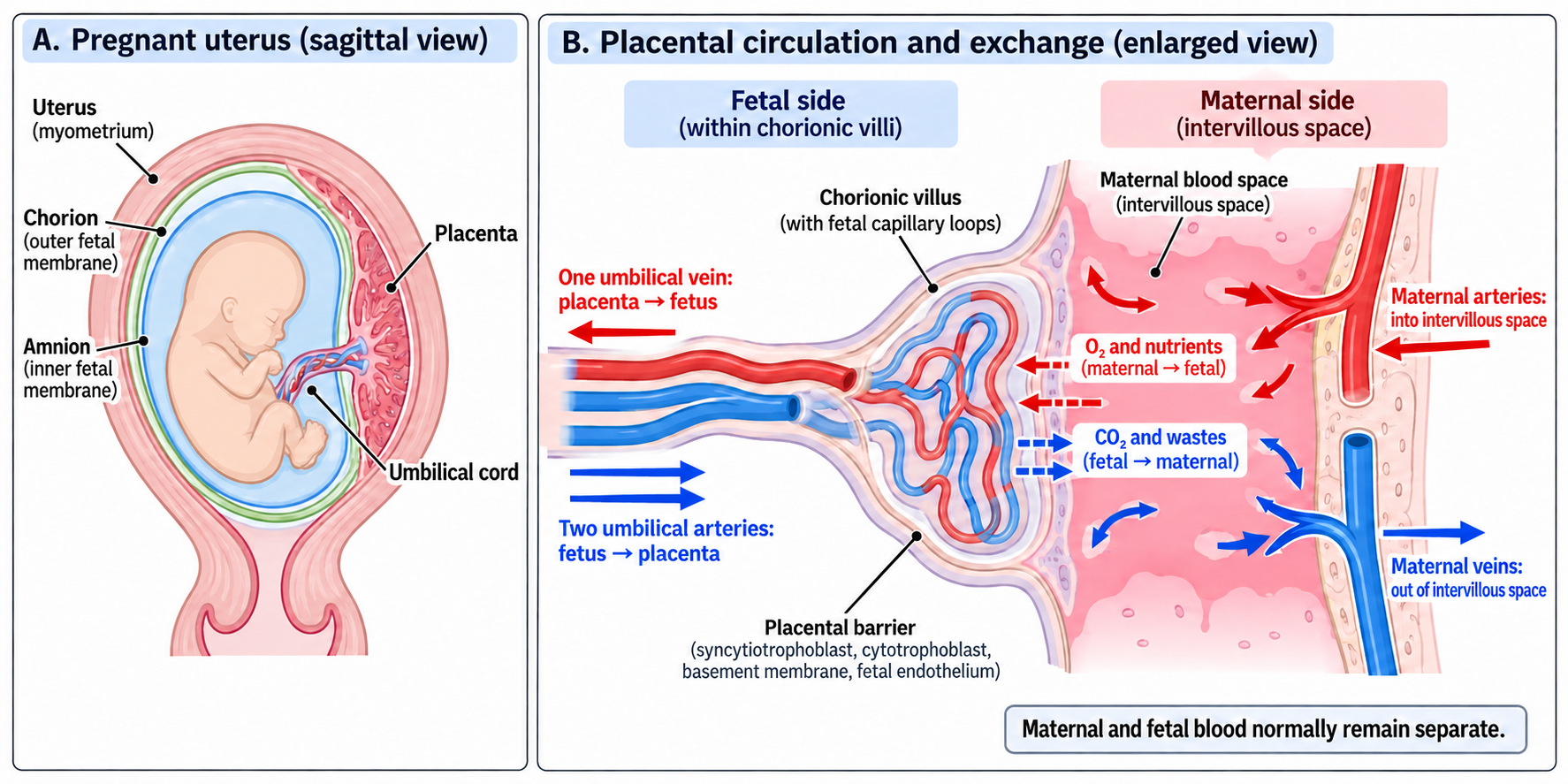
: an organ formed from fetal and maternal tissues that supports nutrient, gas and waste exchange.

1. Placental circulation

: the placenta transfers nutrients and oxygen from maternal blood to fetal blood and removes fetal carbon dioxide and wastes. The two bloodstreams normally remain separate; exchange occurs across the placental barrier.

2. Function of fetal placenta

  • Umbilical vein – carries oxygen- and nutrient-rich blood from the placenta to the fetus
  • Umbilical arteries – carry blood containing carbon dioxide and wastes from the fetus to the placenta

G. Postnatal development

  • Adult hemoglobin gradually replaces fetal hemoglobin during infancy.
  • Lung expansion lowers pulmonary vascular resistance and increases pulmonary blood flow, raising left atrial pressure.
  • Right atrial and pulmonary arterial pressures fall; the higher left atrial pressure promotes functional closure of the foramen ovale.
  • Allometric growth – different growth rates of different body parts.