
VII. Nervous system
Key focus of this chapter: neurons and the nervous system
This chapter reviews neuron structure, neural signaling, and the central and peripheral nervous systems.
A. Organization of the nervous system
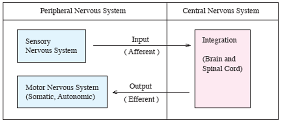
- Afferent
– carry sensory information to the central nervous system.
- Interneurons
– integrate information within the central nervous system.
- Efferent
– carry motor commands from the central nervous system to effectors, such as muscles and glands.
B. Structure of a neuron
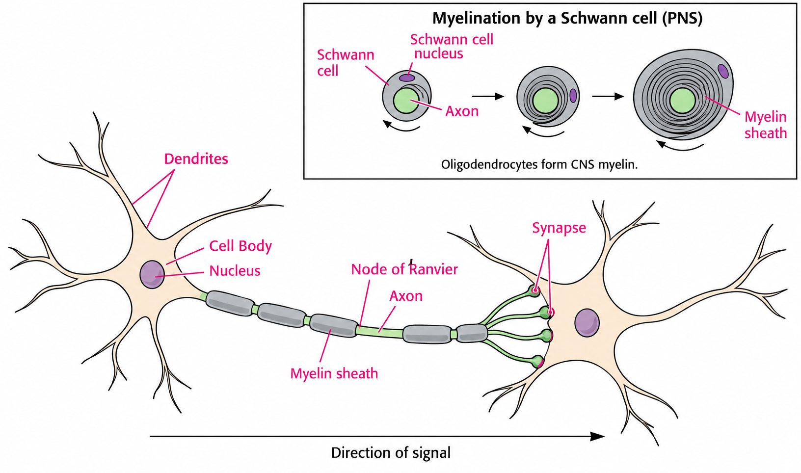
- Dendrites
– receive incoming signals.
– typically highly branched.
- Cell body
– contains the nucleus and carries out cellular metabolism and protein synthesis.
- Axon
– nerve fiber that usually conducts signals away from the cell body toward other cells.
- Myelin sheath
– Schwann cells form myelin around axons in the peripheral nervous system; oligodendrocytes form myelin in the central nervous system.
– the insulating myelin sheath increases the speed of action potential conduction.
** Synapse
: a specialized junction where a neuron communicates with another cell. Chemical synapses usually transmit signals in one direction; electrical synapses use gap junctions and are often bidirectional.
C. Transmission
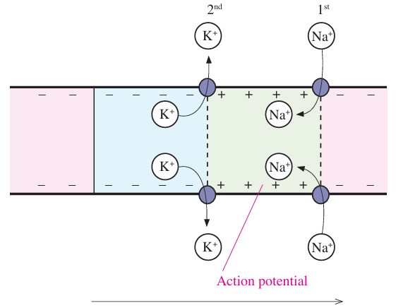
1. Transmission of action potential
- ① Opening of voltage-gated Na+ channels allows Na+ influx and rapid depolarization.
- ② Na+ channel inactivation and opening of voltage-gated K+ channels allow K+ efflux and repolarization.
- ③ Local currents depolarize adjacent membrane to threshold, regenerating the action potential along the axon without loss of amplitude.
2. Action potential with voltage-gated ion channels
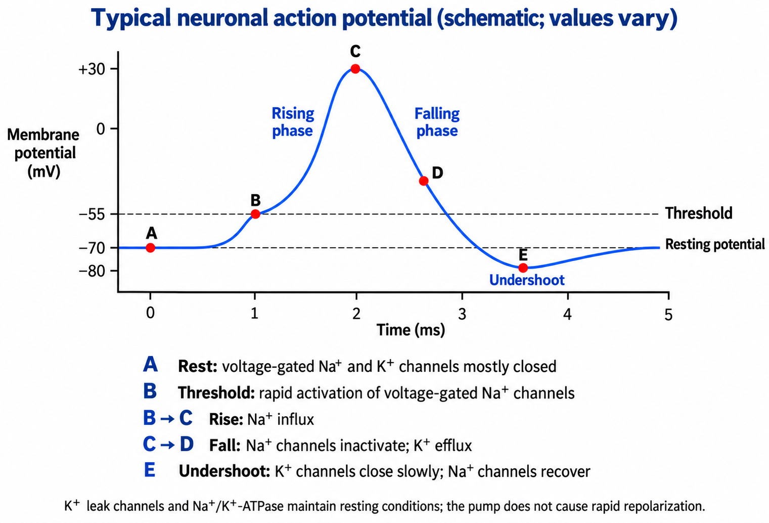
a. Resting state
- At rest, potassium leak channels help establish the membrane potential; most voltage-gated Na+ and K+ channels are closed.
- The interior is negative relative to the outside (often about −70 mV). Na+ concentration is higher outside, and K+ concentration is higher inside; the Na+/K+-ATPase maintains these concentration gradients.
b. Depolarization
- A depolarizing stimulus moves the membrane potential toward threshold.
- At threshold, rapid opening of voltage-gated Na+ channels initiates an all-or-none action potential.
c. Rising phase of action potential
- Na+ influx causes further depolarization and opens additional voltage-gated Na+ channels by positive feedback.
- The membrane potential rises above 0 mV and reaches its peak.
d. Falling phase of action potential
- Voltage-gated Na+ channels inactivate while delayed voltage-gated K+ channels open.
- K+ efflux repolarizes the membrane.
e. Undershoot
- Slow closure of voltage-gated K+ channels briefly makes the membrane more negative than its resting potential.
- During the absolute refractory period, Na+ channel inactivation prevents another action potential. During the relative refractory period, a stronger stimulus is required.
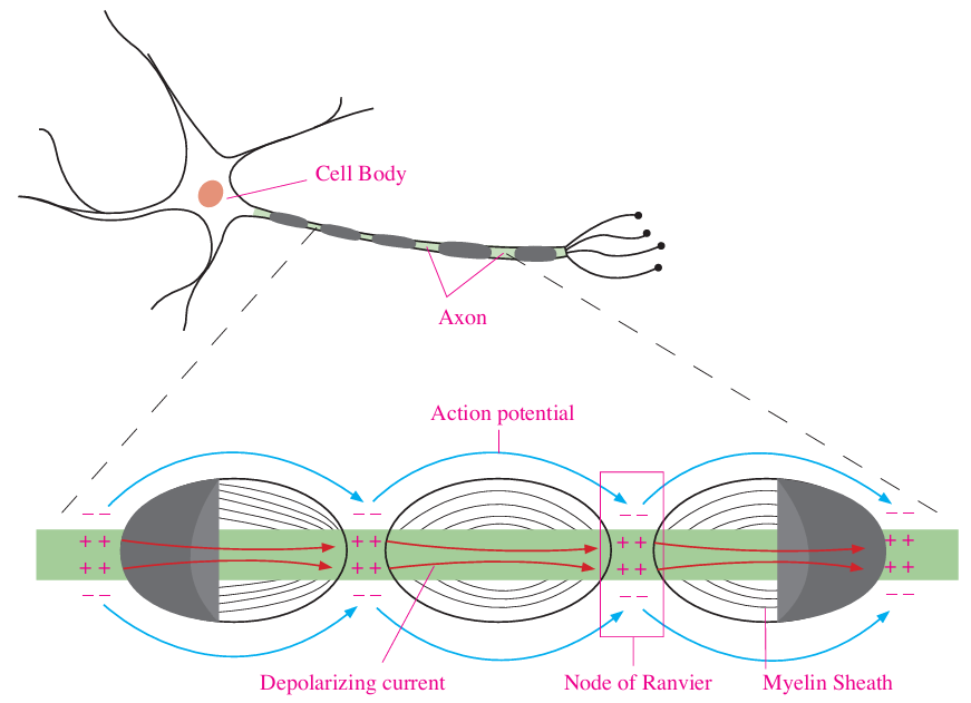
3. Saltatory conduction
: rapid propagation of action potentials along a myelinated axon.
- Action potentials are regenerated at successive nodes of Ranvier, appearing to jump between nodes along the myelinated axon.
- For comparable axons, myelination and a larger axon diameter increase conduction speed.
4. Chemical synapse
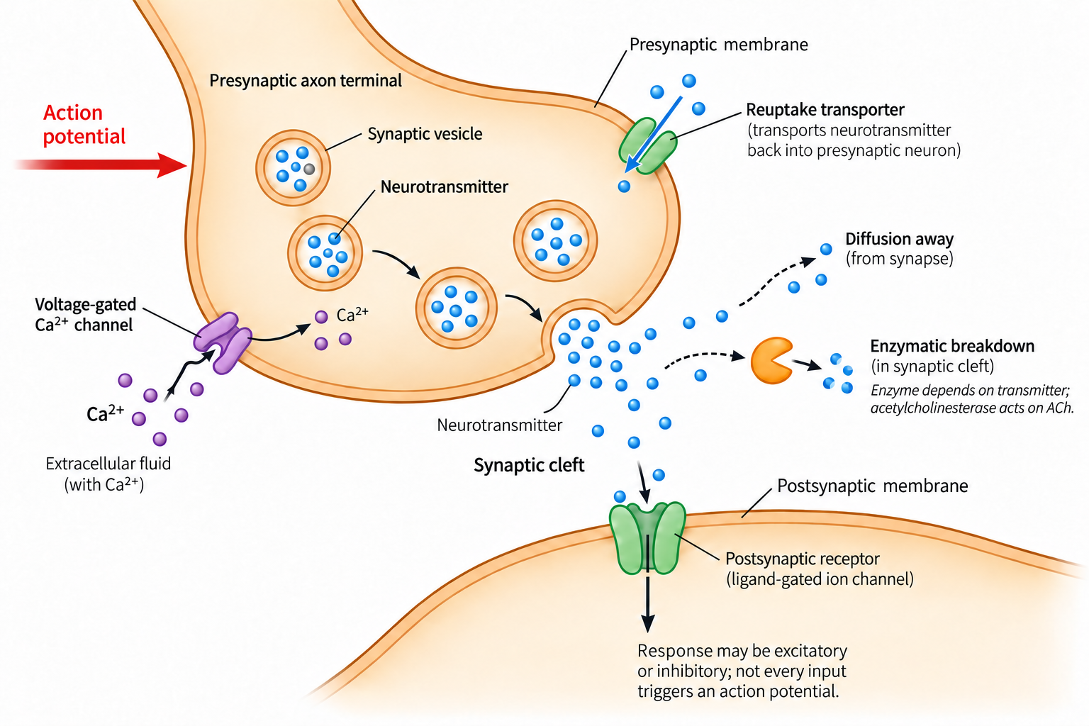
: place for transmission of messages between two neurons or between a neuron and a target cell.
a. Process
- ① An action potential arrives at the presynaptic axon terminal.
- ② The terminal membrane depolarizes.
- ③ Voltage-gated Ca2+ channels open, allowing Ca2+ to enter the terminal.
- ④ Increased cytosolic Ca2+ triggers fusion of neurotransmitter-containing synaptic vesicles with the presynaptic membrane and release by exocytosis.
- ⑤ Neurotransmitter binds postsynaptic receptors, producing excitatory or inhibitory responses. An action potential occurs only if depolarization reaches threshold at the postsynaptic neuron’s trigger zone.
- ⑥ Neurotransmitter is cleared by reuptake, diffusion or enzymatic breakdown; acetylcholinesterase specifically breaks down acetylcholine.
b. Function of each part
- Neurotransmitter
– intercellular chemical messengers.
- Synaptic vesicles
– store neurotransmitters in the presynaptic terminal.
- Voltage-gated Ca2+ channel
– open when an action potential depolarizes the presynaptic terminal, allowing Ca2+ to enter.
- Synaptic cleft
– narrow gap between the presynaptic and postsynaptic membranes.
– neurotransmitter molecules diffuse across the cleft to the postsynaptic cell.
- Ligand-gated ion channels
– postsynaptic receptors that open or close ion channels when a neurotransmitter binds.
D. Central nervous system
1. Brain
Regions | Classification | Functions and features | |
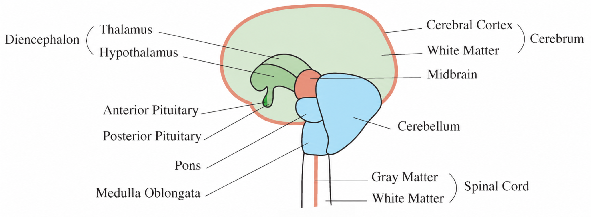
Forebrain | Cerebrum | • Integrating and analyzing sensory information, communication, language, learning, remembering, creative thought, motor response. • Consists of cerebral cortex (gray matter), white matter, and basal nuclei • Gray matter on the outside– dendrite, cell body • Whiter matter on the inside-myelinated axon • Corpus callosum – communication between left brain and right brain | |
Diencephalon | Thalamus | • Sensory input and output for cerebrum • Control of sleeping and wakefulness | |
Hypothalamus | • Regulation of homeostasis such as blood pressure, temperature, or water balance • Sexual drive, thirst, hunger | ||
Midbrain | Midbrain | • Part of brainstem | |
Hindbrain | Pons | • Regulation of breathing rates | |
Medulla oblongata | • Regulation of respiratory system (heart, breathing, blood vessels), swallowing, digestion | ||
Cerebellum | • Coordination, equilibrium (motor balance) | ||
- Brainstem – midbrain, pons, medulla oblongata
- Meninges – protective membranes surrounding the central nervous system
2. Spinal cord
: carries signals between the brain and body and integrates spinal reflexes.
- White matter on the outside – transmission of neural information
- Gray matter on the inside – processing of information
E. Peripheral nervous system
Peripheral nervous system | ||
Autonomic nervous system | Somatic nervous systems | |
• Involuntary control • Afferent (sensor) and efferent (motor) nerves response from internal stimuli such as smooth muscle, cardiac muscles, cardiovascular, digestive organs, excretory, and endocrine. | • Voluntary control (conscious control) by skeletal muscles • Efferent (motor) nerves - response from external stimuli - reflex activity by spinal cord | |
Parasympathetic division | Sympathetic division | |
• Decreasing energy generation to rest and digest states • Respond with decreasing heart rate, respiration, rate of converting glycogen to glucose, and secretion of epinephrine • Increasing digestion • Vagus nerve – one of cranial nerve (starting from the medulla oblongata) to innervate viscera such as stomach, liver, large and small intestines, lung, and heart | • Increasing energy generation from stress • Respond with increasing heart rate, respiration, rate of converting glycogen to glucose, and secretion of epinephrine • Decreasing digestion | |
1. Motor unit
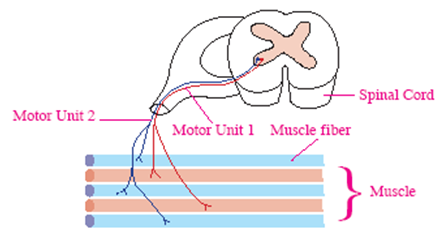
- A motor unit consists of one motor neuron and all the skeletal muscle fibers it innervates.
- One neuron controls many muscle fibers.
2. Autonomic neurotransmitters and receptors
The adrenal medulla receives preganglionic sympathetic ACh at nicotinic receptors and releases epinephrine and norepinephrine into the bloodstream.
Preganglionic neuron | Postganglionic neuron | |||
Neurotransmitter | Receptor | Neurotransmitter | Receptor | |
Parasympathetic system | ACh (acetylcholine) | Nicotinic cholinergic | ACh | Muscarinic cholinergic |
Sympathetic system | ACh | Nicotinic cholinergic | Usually NE (norepinephrine) Exception: ACh to eccrine sweat glands | Usually adrenergic Eccrine sweat glands: muscarinic cholinergic |