X. Immunology

Key focus of this chapter: the immune system

This chapter reviews the immune system, including the lymphatic system, innate defenses, adaptive immunity, and immune responses.

A. Lymphatic system

  • Fluid filtered from blood capillaries enters the interstitial space; excess interstitial fluid then enters lymphatic capillaries as lymph.
  • Lymph nodes filter lymph; macrophages remove microbes and debris, and lymphocytes mount immune responses.
  • Lymph returns through lymphatic vessels and ducts to the venous circulation near the junctions of the subclavian and internal jugular veins.

Lymphatic organs, tissues, and vessels

Functions

Central lymphoid tissues

Thymus

• Supports T-cell maturation and selection

• Produces thymic factors, including thymosins, that support T-cell development

Bone marrow

• Produces blood cells by hematopoiesis

• The primary site of B-cell development and maturation in humans

Peripheral lymphoid tissues and associated vessels

Adenoids

• Lymphoid tissue in the nasopharynx that samples inhaled antigens and supports immune responses, especially during childhood

Tonsils

• Protect against infection

Lymph nodes

• Filter lymph, remove microbes and debris, and provide sites for antigen presentation and lymphocyte activation

Spleen

• Filters blood, removes aged or damaged red blood cells, and mounts immune responses to blood-borne antigens

Peyer’s patch

• Aggregated lymphoid follicles in the ileum; sample intestinal antigens and support mucosal immune responses

Lymphatic vessels

• Collect excess interstitial fluid as lymph and return it to the venous circulation

• Transport immune cells and absorbed dietary lipids

B. Defense system

Immunity

Responses to invading pathogens

Nonspecific

Immunity

(innate Immunity)

External

Defenses

• Skin

• Secretions

• Mucous membranes

Internal Defenses

• Antimicrobial proteins

• Inflammatory responses

• Phagocytic white blood cells

• Natural killer (NK) cells

Specific Immunity
(adaptive Immunity)

• Humoral response (B cells and antibody-secreting plasma cells)

• Cell-mediated response (T cells, including helper and cytotoxic T cells)

C. Nonspecific immunity

: rapid, innate responses that recognize broad patterns associated with microbes or tissue damage.

1. External defenses

a. Skin – forms a physical barrier against invading microbes.

b. Secretions – lysozyme in tears and saliva breaks down peptidoglycan in bacterial cell walls.

c. Mucous membranes – viscous mucus secreted by mucous membranes protects the respiratory and digestive tracts by trapping microbes.

2. Internal Defenses

a. Antimicrobial proteins

  • Complement system

– a set of circulating and cell-associated proteins activated in a cascade.

– promotes inflammation and opsonization; the membrane attack complex can lyse susceptible microbes.

  • Interferon

– signaling proteins; type I interferons released during viral infection induce an antiviral state in nearby cells and help activate immune defenses.

b. Inflammatory Response

  • Histamine – released by mast cells; promotes vasodilation and increased vascular permeability during inflammation.
  • Chemokines – signaling proteins that attract leukocytes toward sites of infection or tissue injury by chemotaxis.

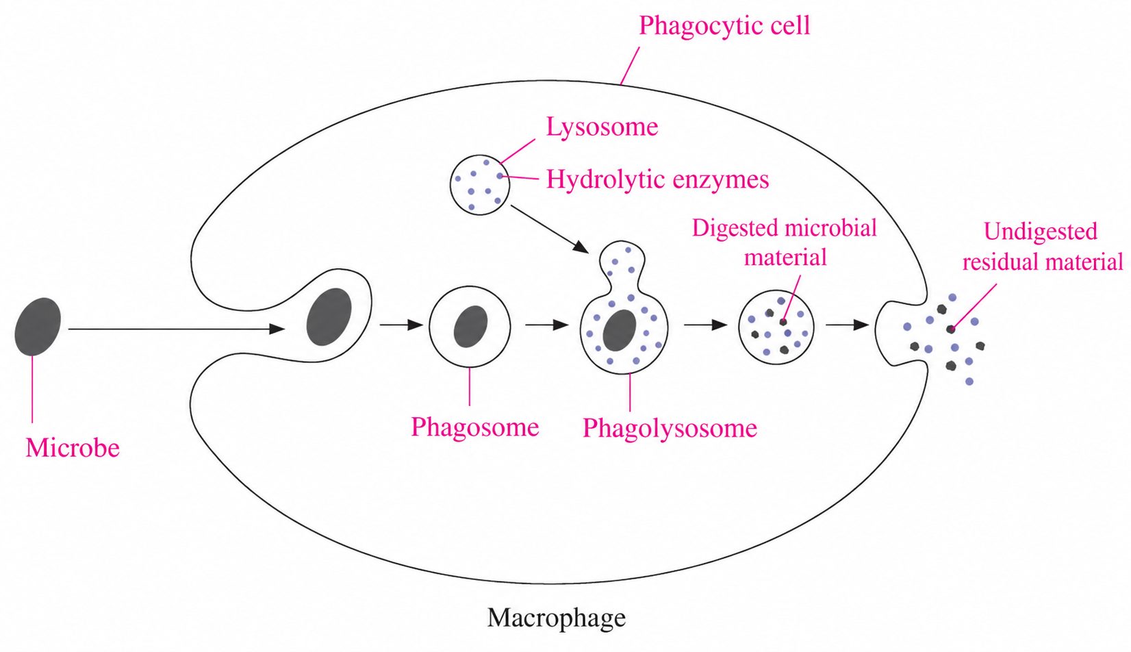
c. Phagocytic cells (macrophage)

Phagocytic white blood cells

Functions and features

Neutrophils

• Rapidly migrate to sites of infection and engulf microbes and foreign particles

Monocytes

• Circulate in blood and can differentiate into macrophages after entering tissues

• Macrophages engulf microbes and debris and can present antigens

Eosinophils

• Have limited phagocytic activity

• Release toxic granule proteins and enzymes that help kill parasites

• Also participate in allergic inflammation; their granule products can damage host tissues

d. Natural killer cells

  • Recognize stressed, infected, or tumor cells without prior antigen-specific sensitization. Perforin helps deliver granzymes, which trigger apoptosis in the target cell.
  • Apoptosis – programmed cell death.

D. Specific immunity

– antigen-specific responses that usually take days to develop after first exposure; memory cells allow a faster, stronger response on reexposure.

1. Basic theory

a. Terminology

  • Antibiotic

– an antibacterial medicine that kills bacteria or inhibits their growth.

– different antibiotic classes target bacterial processes such as cell wall synthesis, protein synthesis, nucleic acid synthesis, or folate metabolism.

– antibiotics do not treat viral infections.

  • Cytokines

– small signaling proteins secreted by immune cells and many other cell types.

– function to help activate lymphocytes and coordinate the immune system against infection.

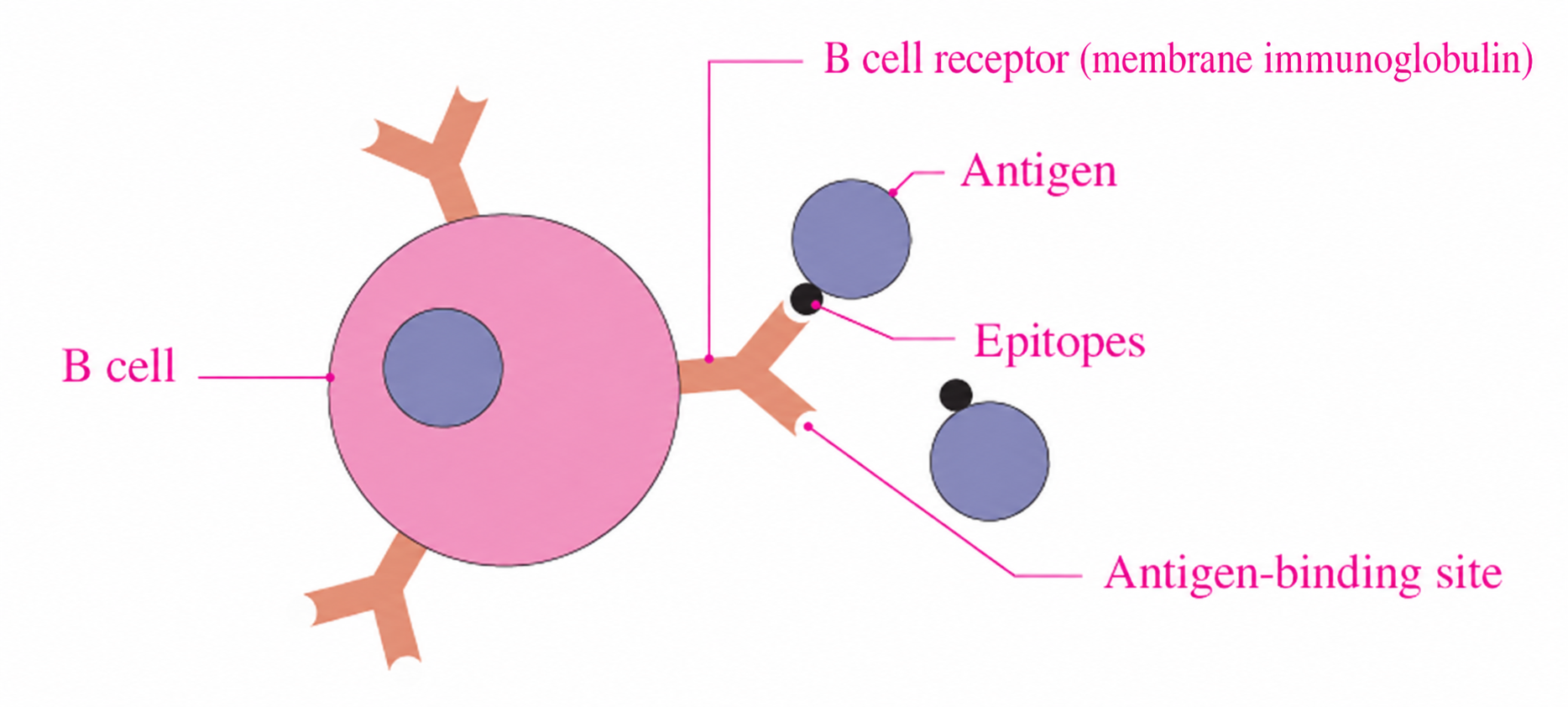
b. Structure of a B cell and an antigen

  • Antibodies

– immunoglobulins secreted by plasma cells (differentiated B cells) and found in blood plasma, interstitial fluid, and secretions.

– bind specific antigenic epitopes; they can neutralize pathogens or toxins and promote clearance through opsonization or complement activation.

  • Antigen

– molecules or molecular structures specifically recognized by antibodies, B cell receptors, or T cell receptors; antigens can be foreign or self-derived.

  • Epitope

– antigenic determinants.

– the specific region of an antigen recognized by an antibody or lymphocyte receptor.

c. Lymphocytes

  • Three main types are B lymphocytes (B cells), T lymphocytes (T cells), and natural killer cells (NK cells). B and T cells mediate adaptive immunity; NK cells function in innate immunity.
  • Lymphocytes are key cells in the body’s immune defenses.

d. Structure of B cell and T cell receptors

Receptors

Features

B cell receptor

• Y-shaped, membrane-bound immunoglobulin with two identical heavy chains and two identical light chains

• Interchain disulfide bonds link the heavy chains to each other and each light chain to a heavy chain; the number varies among immunoglobulin types

• Binds native antigen directly

T cell receptor

• Most T cell receptors consist of two different polypeptide chains, α and β, linked by a disulfide bond

• Recognizes antigenic peptides presented by MHC molecules; a minority of T cells have γδ receptors

e. Lymphocyte development

: lymphocytes arise from hematopoietic stem cells in the bone marrow. In humans, B cells mature in the bone marrow, whereas T-cell precursors migrate to the thymus to mature and undergo selection.

f. Clonal selection of lymphocytes

  • Clonal selection – an antigen activates lymphocytes with matching receptors, together with the required costimulatory signals, leading to clonal expansion and differentiation
  • Effector cells (including plasma cells and effector T cells)

– many are short-lived, but some plasma cells are long-lived.

– act against the antigen that selected the clone; plasma cells secrete antibodies, and effector T cells carry out cellular responses.

  • Memory cells

– long-lived cells.

– retain specificity for the antigen that selected the clone.

  • Primary immune response

– antigen-specific B and T cells proliferate and differentiate; plasma cells secrete antibodies, and effector T cells become active.

– clinical illness may occur, but a primary response does not necessarily cause symptoms.

  • Secondary immune response

– reexposure to the same antigen activates memory cells, usually producing a faster, stronger, and longer-lasting response than the primary response.

2. Humoral and cell-mediated immunities

  • Macrophage

– engulfs microorganisms and presents antigens.

  • Helper T cell

– recognizes peptide–MHC II complexes on antigen-presenting cells, including dendritic cells, macrophages, and B cells.

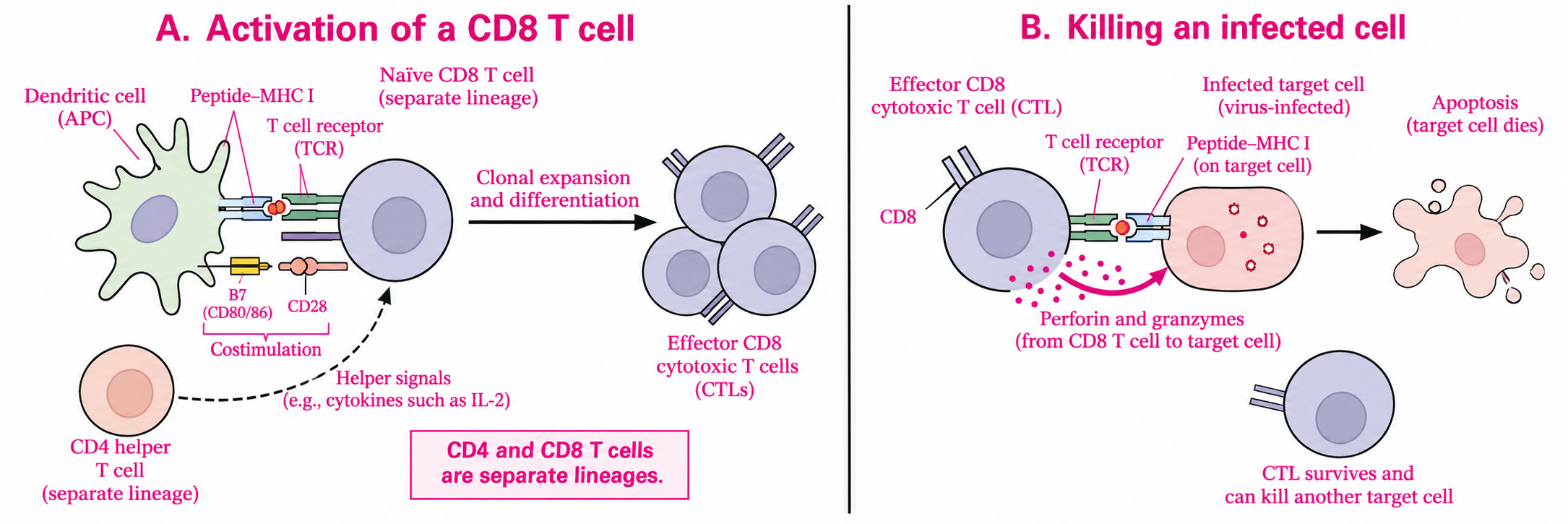
– provides signals that help activate B cells and support CD8 cytotoxic T-cell responses; dendritic cells are the main cells that prime naïve T cells.

a. Humoral immunity

: involves B cells that proliferate and differentiate into antibody-secreting plasma cells and memory B cells. Antibodies defend against extracellular pathogens and toxins in body fluids.

  • B Cells

– play a central role in the humoral immune response.

– bind specific antigens through B cell receptors; responses to most protein antigens also require helper T-cell signals.

– undergo clonal expansion and generate memory B cells.

– differentiate into plasma cells that secrete antibodies into blood, lymph, and other body fluids.

  • Memory B cells
  • Plasma cells – produce antibodies
  • Classes of antibodies

Classes

Shapes

Features

IgA

• Secretory IgA is usually a dimer with a J chain and secretory component; serum IgA is mainly monomeric

• Present in breast milk, mucosal secretions, tears, and saliva

• Provides local mucosal defense and passive protection to nursing infants

IgD

• A membrane-bound antigen receptor coexpressed with IgM on mature naïve B cells

• Participates in B-cell activation

IgE

• Binds mast cells and basophils; mediates immediate allergic reactions

• Contributes to defense against parasitic worms

IgG

• The most abundant antibody class in blood

• Functions in neutralization, agglutination, and opsonization; some subclasses activate complement

• Crosses the placenta and provides passive protection to the fetus and newborn

• Often predominates in secondary antibody responses; can also be produced during primary responses

IgM

• Secreted mainly as a pentamer with a J chain; membrane IgM on B cells is monomeric

• Usually the first antibody class secreted in a primary response

• Efficiently activates complement and agglutinates antigens; also contributes to neutralization

b. Cell-mediated immunity

: involves antigen-specific T-cell responses; cytotoxic T cells proliferate and differentiate into effectors that kill infected or cancer cells.

  • Cytotoxic T cell

– important role in the cell-mediated immune response.

– develops from a separate CD8 T-cell lineage in the thymus, distinct from CD4 helper T cells.

– recognizes peptide–MHC I complexes on infected or cancer cells. Perforin helps deliver granzymes, which trigger apoptosis in the target cell.

  • Class II MHC molecules

– are expressed mainly by professional antigen-presenting cells (dendritic cells, macrophages, and B cells) and present peptides from extracellular antigens to CD4 helper T cells.

  • Class I MHC molecules

– are expressed on nearly all nucleated cells and present peptides from intracellular proteins to CD8 cytotoxic T cells. Mature red blood cells lack MHC I.

  • T cell receptor

– usually recognizes an antigenic peptide bound to an MHC molecule, rather than free antigen.

E. Immune responses

1. Generating immunity

  • Active immunity

– protection produced by the person’s own immune response after antigen exposure; it generates immune memory.

– Examples: infection-induced immunity and vaccination

  • Passive immunity

– temporary protection supplied by preformed antibodies from another source; it acts immediately but does not create immune memory in the recipient.

– Examples: maternal IgG crossing the placenta, secretory IgA in breast milk protecting the infant’s mucosal surfaces, and administered immune globulin.
Rh incompatibility is a different process: in a sensitized Rh− mother, anti-D IgG can cross the placenta and destroy an Rh+ fetus’s red blood cells, causing hemolytic disease of the fetus and newborn. This is harmful antibody-mediated disease, not protective passive immunity.

 2. Blood group

This table shows ABO compatibility for red blood cell transfusions, not plasma transfusions. Rh type, other blood group antigens, and crossmatching must also be considered.

Blood type

Anti-A/anti-B antibodies in recipient plasma

ABO-incompatible donor red cells

ABO-compatible donor red cells

A

Anti-B

B, AB

A, O

B

Anti-A

A, AB

B, O

AB

None

None

A, B, AB, O

O

Anti-A, Anti-B

A, B, AB

O