
VIII. Animal Physiology
Key focus of this chapter: digestion hormones
This chapter focuses on digestion hormones and gives concise summaries of the important things about respiration, cardiovascular system, osmoregulation and excretion, endocrinology, and nutrients.
A. Respiration
: process of gas exchange in the body.
- Internal respiration
– gas exchange between systemic capillary blood and body tissues.
– Cellular respiration is the metabolic production of ATP, distinct from internal respiration.
- External respiration
– gas exchange between alveoli and pulmonary capillary blood.
– O2 diffuses into blood; CO2 diffuses into alveoli.
– Ventilation moves air into and out of the lungs; circulation transports gases between the lungs and tissues.
– Gas exchange at systemic tissues is internal respiration.
- Figure of external respiration

1. Respiratory tract
a. Air Passage


b. Function of each part
Classification | Features and roles |
Pharynx | • Muscular tube for passageway of digestion and respiration |
Larynx | • Cartilaginous voice box that maintains an open airway • Sound generating part • Located between the pharynx and trachea, anterior to the esophagus |
Epiglottis | • Flap tissue to prevent food from entering the respiratory tract |
Trachea | • Rigid structures composed of C-shaped ring of cartilage to keep air flow • Wind pipe |
Bronchi | • Lead air to each lung |
Bronchioles | • Branches of bronchi • Small airways without cartilage; smooth muscle regulates airflow |
Alveoli | • Air sac for gas exchange between lung and capillary |
2. Inspiration and expiration
a. Inspiration
- The diaphragm contracts and moves downward, increasing thoracic volume
- The lungs expand; alveolar pressure falls below atmospheric pressure
- Air moves into alveoli
b. Expiration
- During quiet expiration, the diaphragm relaxes and moves upward
- Elastic recoil decreases lung volume; alveolar pressure rises above atmospheric pressure
- Air moves out from alveoli
B. Cardiovascular System
1. Animal circulatory systems
Animal | Number of Chambers of heart | Number of Circuits |
Fish | Two | One |
Amphibians and most reptiles (lizards, turtles, snakes) | Three | Two |
Birds, mammals, and crocodilians | Four | Two |
2. Heart of mammal
a. The pathway of circulation


b. Anatomy of heart
Classification | Features and roles | |
Atrioventricular valves | • Permit flow from atria to ventricles • Prevent backflow into atrium • Flap composed of connective tissue | |
Semilunar valves | • Permit flow from ventricles into the aorta and pulmonary trunk • Prevent backflow into ventricle • Flap composed of connective tissue | |
Intercalated disks | • Junctional complexes between adjacent cardiac muscle cells, including gap junctions and desmosomes • Gap junctions transmit electrical signals; anchoring junctions mechanically couple cells for coordinated contraction | |
Atrium | • Chamber for receiving blood | |
Ventricle | • Chamber for pumping blood out | |
Conduction system | Sino-atrial node (SA node) | • Functions as pacemaker • Located in the wall of the right atrium • Initiates and controls the rate of heart beat to contract all heart muscles |
Atrioventricular node (AV node) | • Located in the inferior interatrial septum near the tricuspid valve • Delays electrical conduction, allowing ventricular filling before ventricular contraction | |
- Systole – phase of ventricular contraction
- Diastole – phase of ventricular relaxation
- Cardiac output
- Stroke volume
3. Blood vessels
a. Blood circulation in the blood vessels

b. Functions of each part
Blood vessels | Features and functions |
Artery | • Thick muscular wall and large vessel • Transports blood from heart to arteriole • Highest pressure |
Arteriole | • Small vessel • Transports blood from artery to capillary • Branched from artery • Determines the distribution of blood |
Capillary | • Microscopic vessels with a single endothelial layer and basement membrane • Greatest total cross-sectional area and lowest average blood velocity • Connect arterioles to venules within tissues • Exchange gases, nutrients, and wastes with interstitial fluid |
Venule | • Thin and small vessel • Collects blood from capillary • The movements of skeletal muscles help return blood from capillary to heart in venule and vein |
Vein | • Large vessel with low blood pressure • Transports blood from venules to heart • Has valves to prevent back flow |
** Portal venous system
- Transports blood from the capillaries to other capillaries via veins
- e.g., hepatic portal vein
c. Blood pressure
- Main force for the circulatory blood movement
d. Starling forces
- Driving forces for the fluid movement into and out of capillaries
- Capillary hydrostatic pressure and capillary oncotic pressure
Starling forces | Features and functions |
Capillary hydrostatic pressure | • Driving force of fluid from capillary to interstitial fluid. • Usually higher near the arterial end; net movement depends on hydrostatic and oncotic pressure differences and endothelial permeability |
Capillary oncotic pressure (Osmotic pressure) | • Plasma colloid osmotic pressure opposes outward filtration • Mainly generated by plasma proteins such as albumin • Sustained venous reabsorption is not universal; most systemic tissues filter fluid at steady state, with lymphatics returning excess fluid |

4. Blood
a. Cellular elements (45 % of blood)
Cell types | Features and functions | |
Erythrocytes (Red blood cells) | • Have hemoglobin to transport oxygen and carbon dioxide • Mature mammalian erythrocytes lack nuclei and mitochondria | |
Leukocytes (White blood cells) - cells of immune system - have nuclei - larger than red blood cells - can move into the lymphatic system from the blood vessels | Basophils | • Secrete chemicals (histamine and anticoagulant heparin) to allergic reactions or damaged tissues |
Eosinophils | • Minor phagocytic activity • Release enzymes to kill parasites • Granule products can also damage host tissues during inflammation | |
Lymphocytes | • Helper T cells – secrete cytokines to activate B cells and support cytotoxic T cells • B cells – form plasma cells to secrete antibodies • Cytotoxic T cells – secrete molecules (perforin) to kill infected or tumor cells • Natural killer cells (Null cells) - release factors to kill infected or tumor cells | |
Monocytes | • Become macrophages for phagocytic activity | |
Neutrophils | • Phagocytosis of microorganism or foreign particles | |
Platelets | • Hemostasis - process of blood clotting • Lack nuclei | |
b. Plasma elements (55 % of blood)
Classification | Features and functions | |
Ion | • Regulation of osmotic balance, controlling of membrane permeability, pH buffering • e.g., bicarbonate, chloride, sodium, potassium, calcium, magnesium | |
Plasma proteins | Albumin | • Regulation of osmotic balance and pH buffering |
Fibrinogen | • Formation of blood clotting | |
Immunoglobulins | • Antibodies for immune response | |
Water | • Solvent for transporting other materials | |
Other substances | • Nutrients, hormones, waste products | |
c. Transport oxygen
- Hemoglobin
– is a tetrameric protein in red blood cells.
– an allosteric protein.
– each subunit protein has a heme group, which contains iron.
– heme group binds O2 to transport.
– binding affinity of CO is greater than O2
** Myoglobin
– binds O2 to store in muscles.
– similar structure with hemoglobin but with monomer shape.
- Oxygen – binding curve of hemoglobin
– increased O2 binding affinity means increased percent O2 saturation at the same partial pressure of O2
– cooperativity with oxygen binding to hemoglobin.
– Bohr effect means that O2 binding affinity of hemoglobin decreases with low pH (high CO2).

d. Transport carbon dioxide
- Most CO2 (70%) from respiring cells is transported to lungs as bicarbonate ion (HCO3–) mainly in plasma after formation in red blood cells
- Small amount of CO2 (23%) is bound to hemoglobin and transported
- The rest of CO2 (7%) is dissolved in plasma
- Blood pH is buffered by bicarbonate ion (HCO3–)
- Exchanging O2 and CO2 by passive diffusion

e. States of red blood cells

f. Blood clotting
- ① The coagulation cascade forms the factor Xa–Va prothrombinase complex on phospholipid surfaces, with Ca2+, to convert prothrombin to thrombin. Vitamin K supports liver synthesis of functional clotting factors.
- ② Thrombin cleaves soluble fibrinogen to form fibrin.
- ③ Fibrin forms a mesh that stabilizes the platelet plug at the injury site.

C. Digestion
1. Mouth
- Mechanical breakdown of food by teeth.
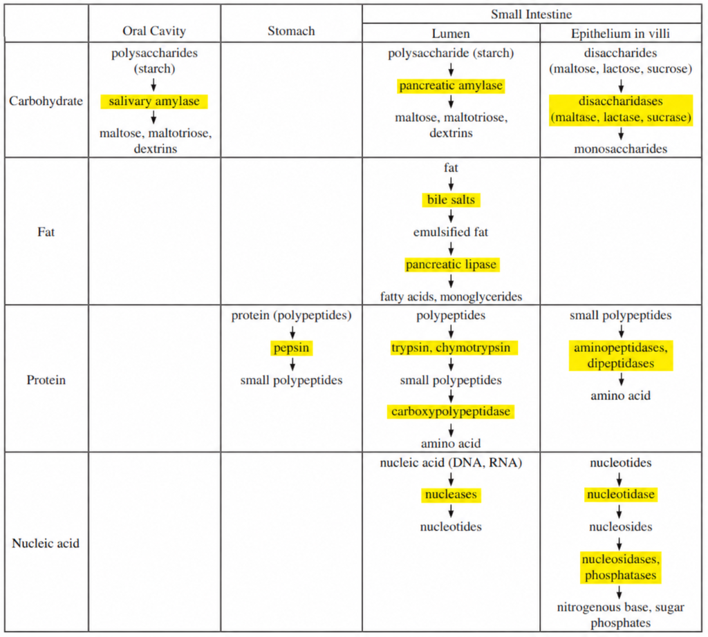
- Chemical breakdown by salivary amylase, which breaks down starch to maltose, maltotriose, and dextrins.
- Moistening of food to form a bolus.
2. Esophagus
- Conducts food from mouth to stomach by waves of muscular contraction called peristalsis.
- Controls the passage of food by muscular ring valves called sphincters.
3. Stomach
- Functions for storage and digestion.
- Mechanical breakdown by churning movements.
- Gastric glands secrete mucus from mucous cells, pepsinogen from chief cells, and hydrochloric acid (HCl) from parietal cells.
- Acid initiates conversion of pepsinogen to pepsin; pepsin activates additional pepsinogen and digests proteins.

- Food mixed with gastric juice becomes acid chyme.
- Mucus lubricates and protects the surface of stomach walls.
- Cardiac orifice (Gastroesophageal sphincter) regulates back flow of food from stomach to esophagus.
- Pyloric sphincter controls the passage of acid chyme from stomach to small intestine.
4. Small intestine
: most chemical digestion for all nutrients occurs in small intestine, which is composed of duodenum, jejunum, and ileum.
a. Secretion to duodenum
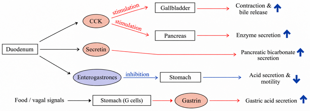
- Pancreas – secretes pancreatic juice, which contains the digestive enzymes and bicarbonate for neutralizing the acid chyme, to duodenum.
- Liver – produces bile; bile salts emulsify fats and aid lipid absorption in the duodenum. The gallbladder stores and concentrates bile.
- Small-intestinal epithelium – provides brush-border enzymes for final carbohydrate and protein digestion.
b. Absorption
- Villi – highly folded mucosal projection to increase surface area for absorption.
- Microvilli – microscopic projection on individual cells.
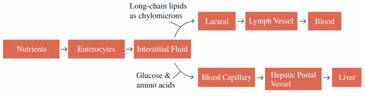
- Long-chain fatty acids and monoglycerides enter enterocytes, are reassembled into triglycerides, and are packaged into chylomicrons that enter lacteals and reach the blood through lymph.
- Glucose and amino acids enter blood capillaries and travel to the liver through the hepatic portal vein.
5. Liver
a. Secretion for digestion
- The liver produces bile; the gallbladder stores and concentrates it.
- Bile contains bile salts and bicarbonate for helping lipid digestion as a function of the emulsifying agent but does not contain enzyme.
b. Multiple functions
- Destruction of aged red blood cells.
- Glycogen synthesis, breakdown, and storage.
- Synthesis of plasma proteins such as albumin, clotting proteins, and angiotensinogen.
- Conversion of ammonia from amino acid metabolism into urea (detoxification).
- Elimination of waste or bacteria from blood.
6. Pancreas
a. Exocrine (secretion of pancreatic juice to duodenum)
- Secrete amylase to digest carbohydrate.
- Secretes trypsinogen; intestinal enteropeptidase activates it to trypsin for protein digestion.
- Secrete lipase to digest fat.
- Secrete bicarbonate ions to neutralize the acid chyme.
b. Endocrine (secretion of hormones into blood stream)
- α cells secrete glucagon to raise blood glucose by promoting hepatic glycogen breakdown and gluconeogenesis.
- β cells secrete insulin to lower blood glucose by promoting uptake in muscle and adipose tissue, promoting storage, and suppressing hepatic glucose production.
7. Large intestine (Colon)
- Composed of four major parts, ascending colon, transverse colon, descending colon, and sigmoid.
- Absorption of water, ions, and other minerals.
- Feces transformation and storage of feces.
- Gut bacteria produce vitamin K (menaquinones), but their contribution to vitamin K status is uncertain; dietary vitamin K is absorbed mainly in the small intestine.
8. Overview of digestion
9. Hormonal control of digestion
D. Osmoregulation and excretion
- Osmoregulation – regulates the concentration of water and solute.
- Excretion – eliminates the nitrogen containing waste product from metabolism.
1. Animal osmoregulation and excretion
Classification of animal | Osmoregulation and excretion | |
Flatworms | • Protonephridia with flame cells | |
Earthworm | • Metanephridia | |
Insect | • Malpighian tubules | |
Bony fish | Freshwater fish | • Excrete abundant dilute urine, conserve salts in kidneys, and actively absorb ions through gills |
Saltwater fish | • Marine bony fish drink seawater, excrete excess NaCl through gills and divalent ions through kidneys, and produce little urine | |
Bird | • Kidneys conserve water; many marine birds use specialized salt glands to excrete excess salt from seawater | |
2. Mammal osmoregulation and excretion
a. Basic renal processes of mammal kidney

- Filtration – hydrostatic pressure drives water and small solutes from glomerular blood into Bowman’s capsule; blood cells and most plasma proteins are retained.
- Secretion – transfer substances such as H+, K+, and drugs from blood into renal tubules, often by active transport.
- Reabsorption – return filtered water and needed solutes from renal tubules to blood by passive or active transport.
- Excretion – elimination of substances in urine; excreted amount = filtered amount − reabsorbed amount + secreted amount.
b. Function of each part
Classification of kidney process | Features and functions | |
Filtration (pressure-driven) | Glomerulus | • Ball shaped capillary tubes • Water and small solutes, including glucose, are freely filtered; most plasma proteins and blood cells are retained |
Bowman’s capsule | • Cup-shaped structure surrounding the glomerulus • Surround glomerulus • Collects the filtrate that passes through the glomerular filtration barrier • Together with the glomerulus, forms the renal corpuscle | |
Secretion and Reabsorption transport) | Proximal tubule | • Reabsorbs filtered glucose and amino acids, most Na⁺ and water; secretes H⁺ and organic substances • Most of the reabsorption of water and salt occurs • pH regulation |
Distal tubule | • Secretion and reabsorption of salt and water • Control K+ and NaCl • pH regulation • Aldosterone increases Na⁺ reabsorption and K⁺ secretion in the late distal tubule and collecting duct | |
Reabsorption transport) | Descending loop of Henle | • Permeable to water; water is reabsorbed into the medulla |
Ascending loop of Henle | • Reabsorbs NaCl and is impermeable to water; the thick ascending limb actively transports salt | |
Excretion | Collecting duct | • Adjusts final urine concentration; ADH increases water reabsorption, and urea recycling supports the medullary gradient |

c. Passage of waste products

E. Endocrinology
1. Hormone
: intercellular chemical signals produced by endocrine glands.
a. Hypothalamus hormones
- Hypothalamic releasing and inhibiting hormones reach the anterior pituitary through portal blood vessels. ADH and oxytocin are synthesized in the hypothalamus and released by the posterior pituitary.
b. Steroid hormones
- Lipid-soluble and poorly soluble in water.
- Cross plasma membranes and bind to intracellular receptors in the cytoplasm or nucleus, usually regulating gene expression.
- e.g., Androgens, Estrogens, Progesterone, cortisol, aldosterone
c. Peptide hormone
- Hydrophilic and dissolved in water.
- Cannot cross plasma membranes and bind to receptors on the surface of a target cell.
- e.g., Most hormones
d. Tropic hormones
- Act on other endocrine glands to stimulate hormone secretion.
- e.g., FSH, LH, TSH, ACTH, hypothalamus hormones
e. Nontropic hormones
- Nontropic hormones act directly on nonendocrine target tissues; e.g., prolactin acts on mammary glands.
2. Hormones of endocrine glands

Hypothalamus Hormones | Pituitary | Hormone | Target | Functions of pituitary hormones |
GHRH (stimulates GH) GHIH (inhibits GH) | Anterior Pituitary | GH (growth hormone) | Liver and bone | • Promotes growth directly and through hepatic IGF-1; affects protein, lipid, and glucose metabolism |
TRH (stimulates TSH) | TSH (thyroid stimulating hormone) | Thyroid | • Stimulates thyroid hormone synthesis and secretion • T3/T4 then act on target tissues to regulate metabolism • Stimulates production of T3 and T4 | |
CRH (stimulates ACTH) | ACTH | Adrenal cortex | • Responds to long term stress • Primarily stimulates glucocorticoid (cortisol) secretion; aldosterone is regulated mainly by RAAS and K⁺ | |
GnRH (stimulates LH and FSH) | LH | Testis and ovary | • Causes the follicle to undergo ovulation • Stimulates the Leydig cells for testosterone secretion in testes | |
FSH (follicle stimulating hormone) | • Promotes ovarian follicle growth and granulosa-cell estrogen production • Stimulates Sertoli cells to support spermatogenesis in testes | |||
TRH (can stimulate prolactin) Dopamine (PIH) (tonic inhibition) | Prolactin | Mammary glands | • Stimulates mammary glands for milk production and secretion | |
MSH | Melanocytes | • Stimulates color changes of skin in reptiles and amphibians | ||
Posterior Pituitary | ADH (antidiuretic hormone, vasopressin) | Kidney tubules | • Increases collecting-duct water permeability and reabsorption; vasoconstriction occurs at higher concentrations • Causes concentrated urine • ADH deficiency or renal resistance to ADH can cause diabetes insipidus | |
Oxytocin | Mammary glands, uterine muscles | • Causes milk ejection by contracting mammary myoepithelial cells • Stimulates uterine contractions during labor |
3. T3 and T4 from Thyroid Gland
: thyroid gland consists of two lobes located on either side of the trachea and controls homeostatic functions and cellular metabolism such as digestion, heart rate, blood pressure, and reproductive functions.
- T3 and T4 are secreted by the thyroid gland, regulated by hypothalamic TRH and anterior pituitary TSH.

- Triiodothyronine (T3)
– has three iodine atoms.
– more powerful activation than T4.
- Thyroxine (T4)
– has four iodine atoms.
– higher rate of production than T3.
– converted to T3 in kidney, liver, or target cells.
- Metabolic disorders of the thyroid gland
Metabolic disorders | Causes and symptoms |
Hyperthyroidism | • Excessive secretion of thyroid hormone • High rate of metabolism with high temperature, poor emotional control, high blood pressure, irritability, profuse sweating, and weight loss • e.g., Graves’ disease; some patients develop thyroid eye disease |
Hypothyroidism | • Insufficient secretion of thyroid hormone • Low rate of metabolism with cold intolerance, lethargy, and weight gain • e.g., myxedema in severe adult hypothyroidism; untreated congenital hypothyroidism can impair growth and neurodevelopment |
4. Regulation of blood calcium levels
a. Parathyroid hormone (PTH)
- Secreted by parathyroid gland when Ca2+ levels in blood are decreased.
- Indirectly promotes osteoclast-mediated bone resorption to increase blood Ca2+.
- Increases renal Ca2+ reabsorption and promotes calcitriol formation, which increases intestinal Ca2+ absorption.
b. Calcitonin
- Secreted by thyroid gland when Ca2+ level in blood is increased.
- Inhibits osteoclast-mediated bone resorption and can increase renal Ca2+ excretion.
- Has a relatively minor role in normal calcium homeostasis in adult humans compared with PTH and vitamin D.

5. Regulation of blood glucose levels
a. Glucagon
- Secreted by alpha cells in pancreas when glucose levels in blood are decreased.
- Promotes hepatic glycogen breakdown and gluconeogenesis.
- Stimulates liver to release glucose into blood stream.
b. Insulin
- Secreted by beta cells in pancreas when glucose levels in blood are increased.
- Promotes glucose uptake in skeletal muscle and adipose tissue and suppresses hepatic glucose production, lowering blood glucose.
- Promotes glycogen synthesis and other nutrient storage processes.

** Islet of Langerhans: composed of Alpha Cells, Beta cells, Delta cells, and F cells in pancreas
6. Stress response
- Adrenal glands lie above the kidneys and contribute to the stress response.

a. Adrenal medulla
- Stimulated by sympathetic preganglionic neurons under hypothalamic control.
- Secretes epinephrine and norepinephrine in response to acute stress.
- Increases blood glucose, heart rate, and blood pressure during the fight-or-flight response.
b. Adrenal cortex
- ACTH stimulates cortisol secretion; aldosterone is regulated mainly by angiotensin II and elevated blood K+.
- The adrenal cortex secretes cortisol (a glucocorticoid) and aldosterone (a mineralocorticoid).
- Cortisol supports stress metabolism, increases glucose availability, and modulates immune and inflammatory responses.
- Aldosterone promotes renal Na+ reabsorption and K+ secretion.
- Na+ retention supports extracellular fluid volume and blood pressure; ADH directly increases collecting-duct water permeability.
7. Other chemical messengers
a. Pheromones
- Chemical substances communicated by odors outside the body.
- Functions in marking territories, courtship behavior, classifying social order between same species.
b. Renin
- Proteolytic enzyme that regulates arterial blood pressure.
- Initiates the renin–angiotensin–aldosterone system by cleaving angiotensinogen to angiotensin I.
c. Melatonin
- An amino acid-derived hormone secreted by the pineal gland in the brain.
- Functions in circadian rhythms.
- Secreted at night.
d. Endorphin
- Neuropeptide of chemical signal.
- Decreases pain perception in brain.
F. Nutrients
1. Vitamin
- Organic compounds
- Needed in small amounts, but problems with deficiencies
- Generally required from the diet in adequate amounts; synthesis varies by species. Humans can synthesize vitamin D in skin and some niacin from tryptophan.
Vitamins | Types | Functions |
Water-soluble Vitamins | Vitamin B1 (thiamine) | • Thiamine pyrophosphate supports oxidative decarboxylation and transketolase; deficiency causes beriberi and Wernicke–Korsakoff syndrome • Contained in grains, peanuts, pork, and legumes |
Vitamin B2 (riboflavin) | • Precursor of FAD (flavin adenine dinucleotide) and FMN (flavin mononucleotide) • Supports redox reactions in energy metabolism; deficiency can cause cheilosis and glossitis • Contained in grains, vegetable, and meats | |
Vitamin B3 (niacin) | • Precursor of the coenzymes NAD⁺ (nicotinamide adenine dinucleotide) and NADP⁺ (nicotinamide adenine dinucleotide phosphate) • Contained in grains, nuts, and meats • Insufficient amount results in pellagra disease | |
Vitamin C (L-ascorbic acid) | • Involved in collagen synthesis • Related with skin of body • Insufficient amount results in scurvy disease • Contained in fruits and vegetables | |
Fat-soluble Vitamins | Vitamin A (retinol) | • Retinal, a vitamin A derivative, is the light-sensitive component of visual pigments • Deficiency can cause night blindness and xerophthalmia • Retinoids occur in animal foods; provitamin A carotenoids occur in dark-green and orange vegetables |
Vitamin D | • Supports intestinal Ca²⁺ and phosphate absorption, bone mineralization, and immune function • Made by skin from sunlight • Contained in egg yolk • Deficiency causes rickets in children and osteomalacia in adults | |
Vitamin E (tocopherol) | • Involved in antioxidant, which is to protect cell membrane against oxidation reaction • Deficiency can cause neuropathy, ataxia, and hemolytic anemia • Contained in nuts, seeds, and vegetable oils | |
Vitamin K (phylloquinone) | • Cofactor for activation of vitamin K-dependent clotting proteins; deficiency can cause bleeding • Dietary vitamin K is absorbed mainly in the small intestine; gut bacteria produce menaquinones, but their contribution is variable • Contained in green vegetables |
2. Minerals
- Inorganic substances
- Needed in small amounts, but cause problems in large amounts
- Phosphorus, magnesium, calcium – important in bone and tooth formation
- Sodium and potassium – function in muscle contraction and nerve conduction
- Iron – binds oxygen in hemoglobin and components of electron carriers