
V. Genetics
Key focus of this chapter: DNA and Chromosome
This chapter focuses on DNA and Chromosome and gives concise summaries of the important things about cell division, mendelian genetics, making protein, and mutation.
A. Cell Division
1. Cell cycle
a. Controlling cell cycle
- G1 checkpoint
– checks DNA damage and whether cell size, nutrients, and growth signals permit entry into S phase; cells may enter the nondividing G0 state.
– cell growth checkpoint
- G2 checkpoint
– checkpoint before M phase.
– checks that DNA replication is complete and DNA damage is repaired before mitosis.
– MPF, a cyclin–Cdk complex, promotes entry into mitosis.
- M checkpoint
– the spindle assembly checkpoint delays anaphase until all chromosomes are properly attached to the spindle through their kinetochores.

b. Mitosis
- Produces genetically identical daughter cells (2n → 2n) in somatic cells.
- Occurs for repairing tissues and growing organisms.
|
Cell phases |
Classification |
Descriptions |
|
|
Interphase |
G1 phase |
• Cell growth • First gap • Duration varies among cell types; G1 is often the longest cell-cycle phase |
|
|
S phase |
• DNA synthesized • Duplicated chromatin |
||
|
G2 phase |
• Cell growth and preparing for mitosis • Second gap |
||
|
M phase |
Mitosis |
Prophase |
• Nuclear envelope breaks down during prometaphase (often grouped with late prophase) • DNA condenses into chromosomes • Spindle microtubule begins to form • Centrosomes move into opposite ends of the cell |
|
Metaphase |
• Chromosomes align along the metaphase plate • Spindle fibers attach to the kinetochores |
||
|
Anaphase |
• Separating and moving sister chromatids to opposite poles by spindle fibers |
||
|
Telophase |
• Nuclear envelopes reappear • Chromosomes begin to be less condensed |
||
|
Cytokinesis |
Cytokinesis |
• Two daughter cells form • Plant cell: forms cell plate • Animal cell: forms cleavage furrow |
|

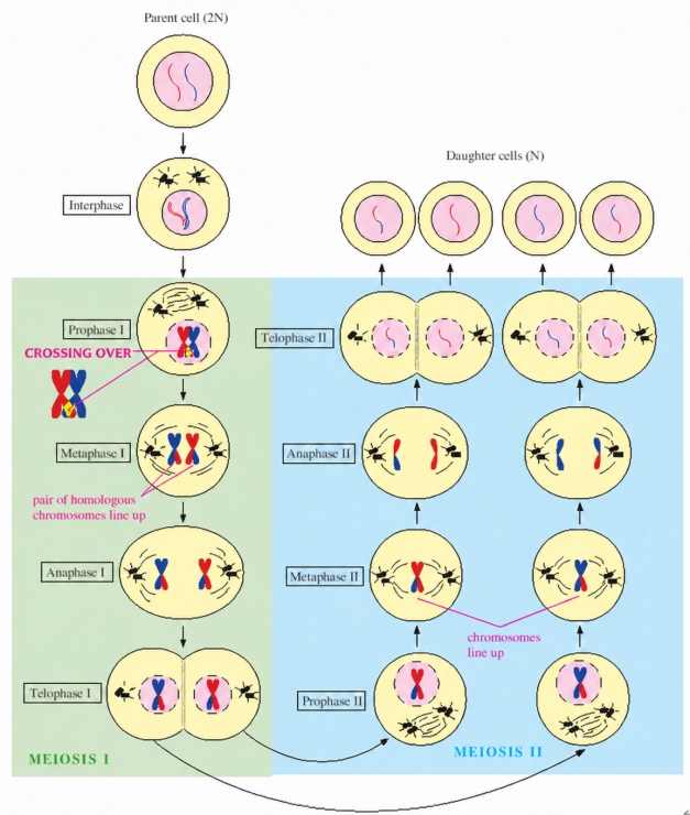
c. Meiosis
- Sexual reproduction resulting in spores or gametes.
- Reducing chromosome sets from diploid to haploid (2n → n) in germ line cells.
- Genetic diversity increases through crossing over between nonsister chromatids of homologous chromosomes in prophase I and independent assortment of homologous chromosome pairs at metaphase I.

** DNA replication occurs during interphase before mitosis and before meiosis I, but not between meiosis I and meiosis II.
Meiosis I | Prophase I | • Nuclear envelope disappears • DNA condenses into chromosomes and then forms homologous chromosome pairs (tetrad) • Synapsis of homologous chromosomes and crossing over between nonsister chromatids; chiasmata mark crossover sites • Spindle microtubule begins to form • Centrosomes move into opposite ends of the cell |
Metaphase I | • Pairs of homologous chromosomes line up along the metaphase plate • Spindle fibers attach to the kinetochores | |
Anaphase I | • Homologous chromosomes separate and move to opposite poles; sister chromatids remain joined. | |
Telophase I & Cytokinesis | • Forming two haploid daughter cells • Plant cell: forming cell plate • Animal cell: forming cleavage furrow | |
Meiosis II | Prophase II | • Spindle microtubule begins to reform • Centrosomes move into opposite ends of the cell |
Metaphase II | • Chromosomes align along the metaphase plate • Spindle fibers attach to the kinetochores | |
Anaphase II | • Sister chromatids separate and move to opposite poles by spindle fibers | |
Telophase II & Cytokinesis | • Nuclear envelopes reappear • Chromosomes begin to be less condensed • Haploid daughter cells form • Cell plate of plant cell and cleavage furrow of animal cell |
2. Cycle of sexual life
- Gametes
– reproductive germ-line cells
– haploid cells (n)
– e.g., Sperm and eggs
- Zygote
– fertilized egg
– diploid cell (2n)
- Somatic cells
– body cells other than cells of the reproductive germ line

B. Population genetics
1. Hardy-Weinberg equilibrium
a. Equation
 |
b. Condition
- Large population
- Isolation (no gene flow)
- No mutation
- Random mating
- No natural selection
2. Genetic Drift
: random changes in allele frequencies from one generation to the next due to chance; effects are strongest in small populations and can reduce genetic variation.
a. Bottleneck effect
- A sharp reduction in population size can cause genetic drift and loss of genetic variation.
b. Founder effect
- A small group establishes a new population; its allele frequencies may differ from those of the original population by chance.
3. Genotype Frequency
a. Inbreeding
- Mating between related individuals increases homozygosity and reduces heterozygosity in offspring.
b. Hybridization
- Crossing genetically different individuals or populations can combine parental traits and increase heterozygosity.
C. Mendelian genetics
1. Terminology
- Allele
– alternative form of a gene.
– e.g., P: dominant allele, p: recessive allele
- Phenotype: observable trait
- Genotype: genetic constitution
- Dominant allele
– under complete dominance, an allele whose associated phenotype is expressed in a heterozygote.
– the same dominant phenotype occurs in dominant homozygotes and heterozygotes.
- Recessive allele
– under complete dominance, the recessive phenotype is masked by the dominant allele in heterozygotes.
– the recessive trait is expressed in recessive homozygotes and can also be expressed in hemizygotes, such as males with an X-linked recessive allele.
- Hemizygous – having only one copy of a gene in a diploid organism; e.g., most X-linked genes in an XY male.
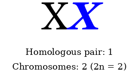
- Pair of homologous chromosomes.
– same staining pattern (same length and centromere position) between two chromosomes.
- Homozygous: A pair of identical alleles.
- Heterozygous: A pair of different alleles.
Pair of homologous chromosomes | Homozygous | Heterozygous |
| AA, aa | Aa |
- Law of segregation
– the two alleles of a gene separate during gamete formation, so each gamete receives one allele.
- Law of independent assortment
– alleles of unlinked genes assort independently during gamete formation; linked genes may not.
- Testcross
– crossing an individual with a dominant phenotype and unknown genotype with a homozygous recessive individual.
– under complete dominance, AA × aa produces all dominant-phenotype offspring; a finite sample of all dominant offspring does not prove the tested parent is AA.
– Aa × aa gives an expected 1:1 dominant:recessive phenotypic ratio; observed ratios vary by chance.
2. Mendelian experiments
Classification | Figures | Features |
P generation |  |  |
F1 generation |  |  |
F2 generation |  |  |
- Monohybrids
– crossing between two heterozygotes of F1 generation having a single characteristic.
– e.g., Bb × Bb → Expected phenotypic ratio of 3:1 under complete dominance
- Dihybrids
– crossing between two heterozygotes of F1 generation having two characteristics.
– e.g., BbDd × BbDd → Expected F2 phenotypic ratio of 9:3:3:1 under complete dominance and independent assortment
3. Law of probability
|
Traits |
Figures |
Features and diseases |
|
Sex-linked traits |
 |
• Sex-linked inheritance involves genes on sex chromosomes. The following examples are X-linked recessive. • Males with a pathogenic allele on their single X chromosome typically express the trait. A carrier mother has a 50% chance of passing the allele to each son. • Females typically express an X-linked recessive trait when both X chromosomes carry pathogenic alleles; heterozygous females may sometimes show signs. Fathers pass their X chromosome to daughters, not sons. • Examples: hemophilia A/B and red–green color blindness |
|
Autosomal traits |
 |
• A pathogenic allele on an autosome can cause a dominant disorder in a heterozygote. Homozygotes may have a more severe phenotype; homozygous achondroplasia is usually lethal. • Examples: achondroplasia, Huntington’s disease, and Hutchinson–Gilford progeria syndrome (typically caused by a de novo variant) |
 |
• An autosomal recessive disorder typically requires pathogenic variants in both copies of a gene, either homozygous or compound heterozygous; heterozygotes are usually carriers. • Examples: sickle cell anemia, phenylketonuria (PKU), oculocutaneous albinism, galactosemia, and Tay–Sachs disease |
4. Pedigrees
- Autosomal traits
– autosomal traits involve genes on autosomes. An affected daughter with an unaffected father alone does not establish autosomal inheritance.

- Dominant traits
– often appear in successive generations, but reduced penetrance or a de novo variant can alter this pattern.

- Recessive traits
– unaffected carrier parents can have an affected child; recessive traits may skip generations.

5. Inheritance
a. Complete dominance
- The heterozygote has the same phenotype as the dominant homozygote.
- Purple flower × white flower → purple flower
b. Incomplete dominance
- The heterozygote has a phenotype intermediate between the two homozygotes.
- e.g., White flower × red flower → pink flower
c. Codominance
- Both alleles are expressed in the heterozygote.
- e.g., roan cattle: heterozygotes have both red and white hairs.
- e.g., ABO blood group has three common alleles: IA, IB, and i (multiple alleles). IA and IB are codominant; both are dominant to i.
Phenotype | Genotype | Antibodies |
A |  | Anti-B |
B |  | Anti-A |
AB |  | None |
O |  | Anti-A, Anti-B |
d. Pleiotropy
- A single gene affects multiple phenotypic traits.
- e.g., Sickle cell disease
e. Epistasis
- One gene masks or modifies the phenotypic effect of a gene at another locus.
- e.g., Color of mice
f. Polygenic inheritance
- Multiple genes contribute to a single trait, often producing continuous variation.
- e.g., Skin color
g. Penetrance
- Percentage of individuals with a specified genotype who express its associated phenotype.
h. Multifactorial disorder
- A disorder influenced by both genetic and environmental factors.
i. Maternal effect
- The offspring’s phenotype is determined by the mother’s nuclear genotype through gene products deposited in the egg. This differs from mitochondrial inheritance, in which mitochondrial DNA is inherited maternally.
D. Gene experiment
1. Griffith transformation experiment
a. Experiment
– 1) injection of living S cells (encapsulated bacteria) into a mouse → mouse dies
– 2) injection of living R cells (nonencapsulated bacteria) into a mouse → mouse lives
– 3) injection of heat-killed S cells into a mouse → mouse lives
– 4) injection of a mixture of heat-killed S cells and living R cells into a mouse → mouse dies
b. Conclusion
- The living R bacteria were converted into the pathogenic S bacteria by an initially unknown transforming factor from the heat-killed S cells.
2. Avery–MacLeod–McCarty experiment
a. Experiment
– 1) mixture of cell-free extract from heat-killed S cells, living R cells, and protease → living S cells
– 2) mixture of cell-free extract from heat-killed S cells, living R cells, and ribonuclease → living S cells
– 3) mixture of cell-free extract from heat-killed S cells, living R cells, and deoxyribonuclease → no living S cells
b. Conclusion
- DNA is the transforming factor.
E. Chromosome basics
1. Chromosomes in humans
- 46 chromosomes in diploid cells (2n).
- 23 chromosomes in haploid gametes (n).
- A typical human diploid somatic cell has 44 autosomes and two sex chromosomes, usually XX or XY.
2. Chromosome appearances
|
Classification |
Features |
|
Metacentric |
Location of centromere at middle of chromosome |
|
Acrocentric |
Location of the centromere near one end of the chromosome |
|
Telocentric |
The centromere is at the end of the chromosome. Telocentric chromosomes are absent from a normal human karyotype. |
3. Level of chromosomal organization

- Histones: proteins around which DNA wraps to form nucleosomes.
This diagram represents a classical hierarchical model of chromatin packaging. In living cells, chromatin organization is heterogeneous and dynamic; regular 30-nm fibers and 300-nm loops are not universal structures.
4. Linked genes
: genes on the same chromosome tend to be inherited together. Crossing over between homologous chromosomes can produce recombinant combinations of alleles.
- Recombination frequency estimates genetic distance between linked genes; 1% recombination corresponds to about 1 centimorgan for short distances.
- Recombination frequency generally increases with genetic distance and has a theoretical maximum of 50%. Multiple crossovers can cause it to underestimate longer map distances.
- Linkage map

5. Sex-linked genes
: genes on X or Y chromosomes.
- X-linked recessive traits
– hemophilia
– color blindness
- Barr body: an inactivated X chromosome
6. Chromosomal alteration
a. Changing chromosome number

- Nondisjunction
– failure of homologous chromosomes to separate in meiosis I or sister chromatids to separate in meiosis II or mitosis.
- Aneuploidy
: gain or loss of individual chromosomes, often caused by nondisjunction during meiosis or mitosis.
– monosomy (2n−1): one copy of a particular chromosome instead of the usual two.
– trisomy (2n+1): three copies of a particular chromosome instead of the usual two.
- Polyploid
: more than two sets of chromosomes.
– triploidy (3n)
– tetraploid (4n)
b. Changing chromosomal structures
- Translocation
– transfer of a chromosome segment to a nonhomologous chromosome; reciprocal translocations exchange segments.
- Deletion
– lost chromosomal segment.
- Duplication
– repeating chromosomal segment.
- Inversion
– reverse attachment of chromosomal segment.

7. Human disorders
a. Down syndrome
- Usually caused by trisomy 21 from nondisjunction; translocation and mosaic forms also occur.
- Typically 47 chromosomes in full trisomy 21.
b. Klinefelter syndrome
- An extra X chromosome, usually resulting from sex-chromosome nondisjunction.
- Typically an XXY male with reduced testosterone and infertility; mosaic forms occur.
- May include reduced facial and body hair and gynecomastia.
- Typically 47 chromosomes (47,XXY).
c. Turner syndrome
- One X chromosome is missing or structurally altered; mosaic forms also occur.
- Typically a female with ovarian insufficiency and infertility, although some individuals can conceive.
- Monosomy X in the classic form.
- 45 chromosomes (45,X) in the classic form.
F. DNA replication
1. DNA structure
- Rosalind Franklin made X-ray diffraction photograph.
- James Watson and Francis Crick proposed the double-helix model of DNA.
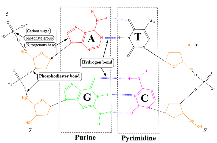
- DNA backbone: deoxyribose sugars linked by phosphate groups.
- Purine: A (adenine), G (guanine).
- Pyrimidine: C (cytosine), T (thymine).
- Two hydrogen bonds between A and T, three hydrogen bonds between G and C.
- For DNA of similar length under the same conditions, higher GC content generally increases the melting temperature.
2. Chargaff’s rules
- In double-stranded DNA: A = T, G = C, and A + G = C + T
- e.g., A = 30%, G = 20%, C = 20%, T = 30%
3. Semiconservative model
- The two parental DNA strands separate, and each serves as a template for a new complementary strand. Each daughter DNA molecule contains one parental strand and one newly synthesized strand.
- If only parental DNA is radiolabeled and new nucleotides are unlabeled, the fraction of labeled strands falls to 50% after one round and 25% after two rounds of replication. Both first-generation daughter molecules contain a labeled parental strand; after two rounds, two of four molecules do.
- The total parental radiolabel is retained if labeled DNA is not lost or degraded and radioactive decay is negligible or corrected for; it is diluted among increasing amounts of DNA, not halved by replication.
- In the figure below, “50% strands parental” and “25% strands parental” refer to the fraction of labeled strands under these assumptions. “Conservative” means both original strands remain together in one molecule; the other molecule has two new strands.

4. DNA replication
|
DNA replication |
Transcription (DNA template → RNA) |
|
A → T; T → A G → C; C → G |
A → U; T → A G → C; C → G |
a. Origins of replication
- Replication fork
– Y shaped site by helicases.
– places of elongation of new strand DNA.
b. Elongation
- Leading strand
– DNA is synthesized continuously in the 5′→3′ direction (by DNA Pol III in E. coli).
– synthesis proceeds toward the advancing replication fork.
- Lagging strand (Okazaki fragments)
– each Okazaki fragment is synthesized in the 5′→3′ direction (by DNA Pol III in E. coli).
– each fragment is synthesized away from the advancing replication fork.
– both leading and lagging strands synthesize DNA only 5′→3′; lagging-strand synthesis is discontinuous, not 3′→5′ DNA synthesis.
c. Enzymes for DNA synthesis
- Topoisomerase
– relieving the strain of tighter twisting from untwisting of parental DNA.
- Helicase: Separating the parental double strands of DNA
- Single-strand binding protein
– binding and stabilizing unwound single-stranded DNA, preventing the strands from reannealing.
- Primase
– synthesizing short RNA primers on the DNA template to provide a 3′-OH starting point for DNA polymerase.
- DNA polymerase
– in E. coli, DNA Pol III is the main enzyme that synthesizes new DNA.
– in E. coli, DNA Pol I removes RNA primers and replaces them with DNA.
– in eukaryotes, Pol α–primase initiates synthesis; Pol ε primarily synthesizes the leading strand and Pol δ primarily synthesizes the lagging strand. RNase H and FEN1 participate in RNA-primer removal. Replicative polymerases such as Pol III, Pol δ, and Pol ε proofread DNA.
- DNA ligase
– joining the ends of new strand of DNA.
- Telomerase
– extending chromosome ends using an internal RNA template; in humans, it adds TTAGGG repeats to help counteract telomere shortening.
d. Repairing DNA

G. Making protein
1. Central dogma

2. Transcription
a. Initiation
① A promoter is a DNA region that helps recruit transcription machinery. Some eukaryotic RNA polymerase II promoters contain a TATA box, recognized by the TATA-binding protein; many promoters lack a TATA box.
② General transcription factors and RNA polymerase II assemble at the promoter of a protein-coding gene. Bacterial promoter recognition instead involves a sigma factor associated with RNA polymerase.
③ RNA polymerase reads the DNA template 3′→5′ and synthesizes RNA 5′→3′. In eukaryotes, protein-coding transcripts are initially made as pre-mRNA.
b. Elongation
- RNA polymerase elongates pre-mRNA in the 5′→3′ direction
c. Termination
- Finishing pre-mRNA transcript
3. RNA processing
a. Modification of pre-mRNA ends
- Adding a 5′ cap and a poly(A) tail at the 3′ end of pre-mRNA
b. RNA splicing
: spliceosome removes introns from pre-mRNA and splices exons together to make mRNA.
- Introns: sequences removed from a pre-mRNA during splicing.
- Exons: sequences retained in mature RNA after splicing; they may include protein-coding sequences and untranslated regions (UTRs).
- Spliceosome: snRNPs + other proteins
4. Translation
a. Codons and anticodons
- Codons: three-nucleotide sequences in mRNA specifying an amino acid or a stop signal
– start codon: AUG (met)
– stop codon: UAA, UAG, UGA
- Anticodons: three-nucleotide sequences in the anticodon loop of tRNA that pair antiparallel with complementary mRNA codons
- Wobble: flexible base pairing between the third nucleotide of an mRNA codon and the first nucleotide of the tRNA anticodon (written 5′→3′) allows one tRNA to recognize more than one synonymous codon.
b. tRNA
- Features
– intramolecular base pairing forms short double-stranded stems within a single tRNA molecule.
– presence of modified nucleosides, such as inosine, pseudouridine, and 4-thiouridine.
- Aminoacyl-tRNA synthetase binds an amino acid to 3′ end of tRNA
c. Ribosomes
: making polypeptide after reading mRNA from the 5′ end to the 3′ end and requiring energy (GTP → GDP).
- A site
– aminoacyl-tRNA binding site.
- P site
– holds the peptidyl-tRNA before peptide-bond formation.
– the growing peptide is transferred from the P-site tRNA to the amino acid on the A-site tRNA. During translocation, tRNAs move A→P and P→E.
- E site
– exit of tRNA site.
H. Mutation
1. Point mutation
Point mutation: a genetic change affecting a single nucleotide or nucleotide pair. It may involve a substitution or, under a broader definition, the insertion or deletion of a single nucleotide or nucleotide pair.

- Missense: result of a different amino acid by altering codon
- Nonsense: result of stop codon
- Silent: result of same amino acid although altering codon
2. Frameshift
: insertion or deletion of nucleotides in a coding sequence in a number not divisible by three, shifting the reading frame.
- Can cause serious genetic diseases
- Deletion or insertion

3. Cancer
- Oncogenes: activated or overexpressed forms of proto-oncogenes that can promote uncontrolled cell proliferation.
- Cancer usually develops through multiple cooperating genetic and epigenetic changes; a single somatic mutation does not necessarily cause a tumor.
I. Gene technology
1. Polymerase chain reaction (PCR)
- Rapidly amplifies a specific DNA segment through repeated cycles of denaturation, primer annealing, and extension.
- Useful for critical forensic analysis.
2. X-ray diffraction
- Method to know the three-dimensional molecular structure.
- X-ray crystallography.
3. DNA fingerprint
- Identification of an individual’s DNA by their DNA profiles.
4. Transgenic organism
- An organism whose genome contains foreign DNA introduced artificially, typically from another species.