I. Cell
Key focus of this chapter: biological macromolecules
This chapter focuses on biological macromolecules and gives concise summaries of the important things about cell membrane and cellular respiration in more detail.
A. Biological Macromolecules
Most biological molecules are composed primarily of carbon (C), hydrogen (H), oxygen (O), nitrogen (N), phosphorus (P), and sulfur (S), collectively known as CHNOPS.
• Nucleic acids: C, H, O, N, and P
• Proteins: primarily C, H, O, and N; some proteins also contain S
1. Carbohydrates (Sugars)
- Composed primarily of carbon, hydrogen, and oxygen, often in an approximate 1:2:1 ratio
- Functions: energy source, energy storage, structural support, and cell recognition
Carbohydrates (sugar) | Classification | Description |
Monosaccharide | Glucose | • A six-carbon aldose sugar; the primary energy source for most cells |
Ribose | • A five-carbon sugar found in RNA | |
Deoxyribose | • A five-carbon sugar found in DNA | |
Galactose | • A component of lactose | |
Fructose | • A six-carbon ketose sugar found in many fruits; a component of sucrose | |
Disaccharide | Sucrose | • Glucose + fructose • α(1→2)β glycosidic linkage |
Maltose | • Glucose + glucose • α(1→4) glycosidic linkage | |
Lactose | • Glucose + galactose • β(1→4) glycosidic linkage | |
Polysaccharide | Starch | • A glucose-storage polysaccharide in plants • Composed of amylose and amylopectin • Contains α(1→4) linkages and α(1→6) branch points |
Glycogen | • A highly branched glucose-storage polysaccharide in animals and fungi • Contains α(1→4) linkages and α(1→6) branch points • More highly branched than starch | |
Cellulose | • The primary structural polysaccharide of plant cell walls • Composed of β-glucose monomers joined by β(1→4) glycosidic linkages • Parallel cellulose chains are linked by hydrogen bonds to form microfibrils | |
Chitin | • A structural polysaccharide composed of N-acetylglucosamine (NAG) monomers • A major component of fungal cell walls and arthropod exoskeletons |
2. Proteins
a. Amino acids
Amino acids are the monomers of proteins.
Each amino acid contains a central α-carbon bonded to an amino group, a carboxyl group, a hydrogen atom, and a variable side chain (R group).
The 20 common amino acids differ in the chemical properties of their R groups.
b. Polypeptides
Amino acids are joined by peptide bonds through dehydration (condensation) reactions to form polypeptide chains.
Peptide bonds are formed by dehydration synthesis and broken by hydrolysis.
Functions of proteins include enzymatic catalysis, signaling, regulation, transport, storage, defense, structural support, and movement.
High temperatures, extreme pH conditions, or certain chemicals can denature proteins by disrupting their secondary, tertiary, or quaternary structure, often causing loss of biological function.
Denaturation usually does not break the peptide bonds of the primary structure.
Structures | Features |
Primary (1º) structure | • Unique linear amino acid sequence; peptide bonds. |
Secondary (2º) structure | • α-helices and β-pleated sheets; stabilized by hydrogen bonds between backbone atoms. |
Tertiary (3º) structure | • Overall 3D shape of a single polypeptide; stabilized by hydrogen bonds, ionic interactions, disulfide bonds, van der Waals forces, and hydrophobic interactions among R groups. |
Quaternary (4º) structure | • Association of two or more polypeptide subunits into one functional protein; Example: hemoglobin. |
3. Lipids
a. Fats
Unsaturated fatty acids contain one or more carbon–carbon double bonds.
Cis double bonds create kinks in hydrocarbon tails, preventing tight packing and increasing membrane fluidity.
Unsaturated fats are commonly liquid at room temperature (e.g., many plant oils).
Saturated fatty acids contain no carbon–carbon double bonds.
Their straight hydrocarbon tails pack tightly together, decreasing membrane fluidity.
Saturated fats are commonly solid at room temperature (e.g., butter).
Fats are more energy-dense than carbohydrates and proteins.
A triacylglycerol (triglyceride) consists of one glycerol molecule joined to three fatty acids by ester linkages.
b. Phospholipids
Phospholipids are amphipathic molecules with hydrophilic heads and hydrophobic fatty-acid tails.
They spontaneously form a phospholipid bilayer, the basic structure of cell membranes.
c. Steroids
Steroids are lipids characterized by four fused carbon rings.
Examples include cholesterol, testosterone, estradiol, cortisol, and vitamin D.
Cholesterol is an essential component of animal cell membranes and helps regulate membrane fluidity and permeability.
Cholesterol is also a precursor of steroid hormones, bile acids, and vitamin D.
Waxes are hydrophobic lipids that provide protective, water-resistant coatings.
4. Nucleic acids
Nucleic acids are biological macromolecules that store and transmit genetic information.
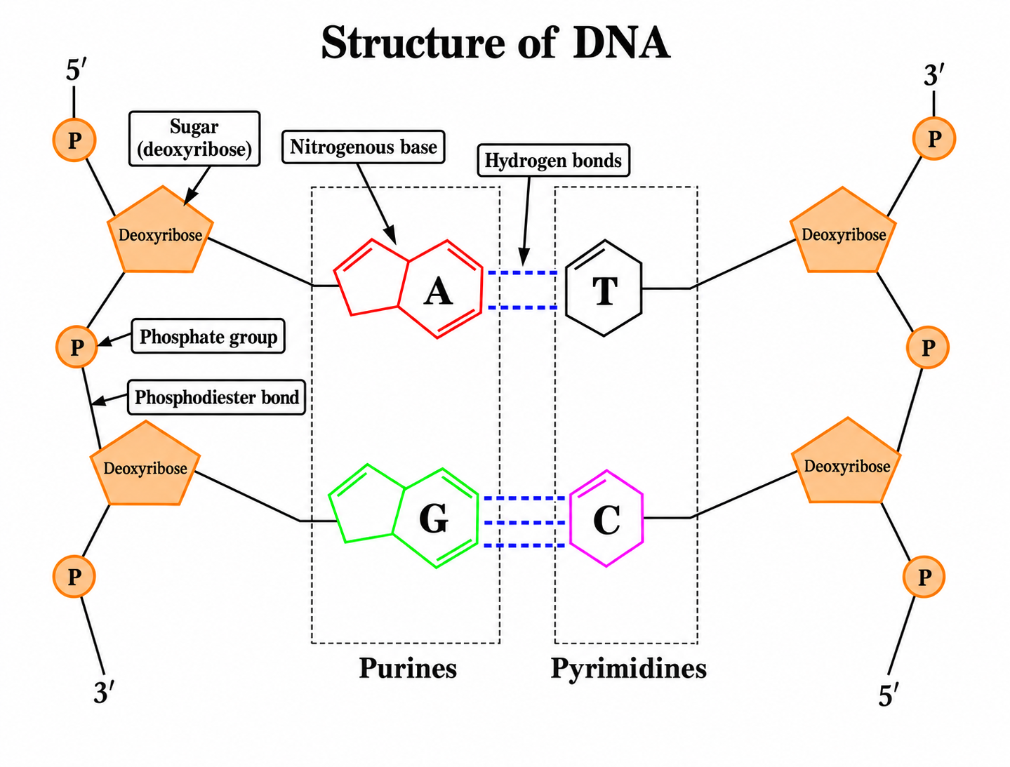
Each nucleotide consists of a five-carbon sugar, a nitrogenous base, and a phosphate group.
Adjacent nucleotides are linked by phosphodiester bonds to form a sugar-phosphate backbone.
In DNA, two antiparallel strands are held together by hydrogen bonds between complementary bases.
Classification | Description |
Nucleoside | Five-carbon sugar + nitrogenous base |
Nucleotide | Five-carbon sugar + nitrogenous base + phosphate group |
Nucleic acids | Polymer of nucleotides linked by phosphodiester bonds |
DNA | RNA | |
Sugar | Deoxyribose | Ribose |
Bases | A, G, C, T | A, G, C, U |
Structure | Usually double-stranded | Usually single-stranded |
Bonds | Hydrogen bonds (between strands) and phosphodiester bonds | Phosphodiester bonds |
B. Shapes and functions of the cells
1. Cell theory
- The cell is the smallest unit of life.
- Cells are the basic structural and functional units of all living organisms.
- All cells arise from preexisting cells.
2. The differences between prokaryotic and eukaryotic Cells
Prokaryotic cell | Eukaryotic cell |
• Cell division by binary fission • Can exchange genetic material by conjugation • Nucleoid (lacks a nuclear envelope) | • Mitotic and meiotic division • Generally larger than prokaryotic cells • Nucleus (w/ nuclear envelope) • Organelles with membrane |
3. Structure of prokaryotic and eukaryotic cells

Structures | Prokaryote | Eukaryote | |
Plant | Animal | ||
Cell wall | Many bacteria have peptidoglycan cell walls; archaeal cell walls lack peptidoglycan. | Existence (cellulose) | None |
Cytoplasm | Existence | Existence | Existence |
Ribosome | Existence | Existence | Existence |
Cytoskeleton | Existence | Existence | Existence |
Nucleoid | Existence | None | None |
Nucleus | None | Existence | Existence |
Mitochondria | None | Existence | Existence |
ER, Golgi | None | Existence | Existence |
Chloroplast | None | Present in photosynthetic plant cells | None |
Vacuole | None | Existence (major) | Existence (minor) |
Centriole | None | Usually absent in typical flowering-plant somatic cells | Usually present in typical animal cells |
Lysosome | None | Lytic vacuoles perform comparable digestive functions | Typically present in animal cells |
a. The functions of organelles
Organelles | Functions and features |
Flagella | • Cell locomotion |
Nucleus | • DNA replication and transcription • Stores genetic information |
Ribosomes | • Site of protein synthesis. • Ribosomal subunits are assembled in the nucleolus. • Functional ribosomes are found in the cytoplasm, mitochondria, and chloroplasts. • Free in the cytoplasm or bound to the rough endoplasmic reticulum (ER). • Composed of rRNA and proteins. |
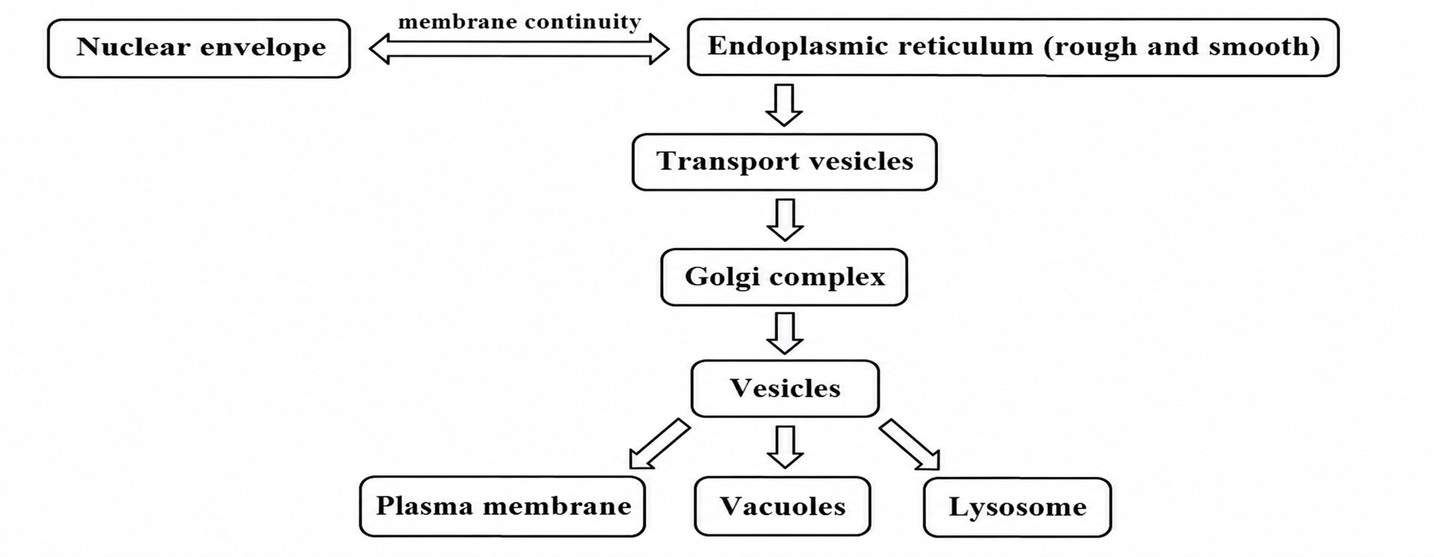
Rough ER | • Studded with ribosomes. • Synthesizes secretory proteins, membrane proteins, and membrane components. |
Smooth ER | • Lacks ribosomes • Lipid synthesis (including steroid hormones) • Calcium storage • Carbohydrate metabolism • Detoxification |
Golgi Apparatus | • Receives proteins and lipids from the ER. • Modifies, sorts, and packages proteins and lipids. • Ships vesicles from the trans face to the plasma membrane or other destinations. |
Lysosomes | • Membrane-bound organelles containing hydrolytic enzymes. • Formed through the Golgi apparatus. • Primarily found in animal cells. |
Peroxisomes | • Contains oxidative enzymes that produce and break down H₂O₂. • Oxidizes fatty acids and detoxifies harmful compounds. • Formed from the ER. |
Vacuole | • Stores nutrients and waste products. • Isolates harmful substances. • Maintains internal pH and turgor pressure. • Surrounded by the tonoplast membrane. |
Mitochondria | • Site of aerobic cellular respiration and ATP production. • Contains its own ribosomes and DNA. • Double membrane, cristae, and matrix. • Originated through endosymbiosis. |
Chloroplasts | • Site of photosynthesis. • Contains its own ribosomes and DNA. • Double membrane, stroma, thylakoids, and chlorophyll. • Originated through endosymbiosis. |
Cytosol | • Main site of many metabolic reactions, including glycolysis. |
Centriole | • Organizes microtubules • Helps form the mitotic spindle during cell division • Found primarily in animal cells |
b. Cytoskeletons
Cytoskeletons | Functions and features |
Microtubules | • Hollow tubes composed of tubulin • Form the mitotic spindle during cell division • Most motile eukaryotic cilia and flagella have a 9+2 microtubule arrangement; primary cilia usually have a 9+0 arrangement • Major component of centrosomes and centrioles • Form cilia and flagella (with dynein for movement) • Serve as tracks for intracellular transport by kinesin and dynein |
Microfilaments | • Cleavage furrow in cytokinesis • Cell crawling (amoeboid movement) • Support the cell cortex and microvilli • Interact with myosin during muscle contraction |
Intermediate filaments | • Tensile strength • Nuclear lamina • Keratin provides mechanical strength to epithelial cells. |
c. Different layers of membranes
Membranes | Organelles |
No membrane | Centriole, Ribosome |
Single membrane | Peroxisome, Lysosome, ER, Golgi apparatus, Vacuole |
Double membrane | Chloroplast, Mitochondria, Nucleus |
d. Extracellular matrix
• Collagen is a major fibrous structural protein of the animal extracellular matrix. Its molecules form a triple helix.
e. Endomembrane system
f. Growth of cell
If a cell’s linear dimensions increase tenfold:
- Area: 102 increase
- Volume: 103 increase
C. Cell communication
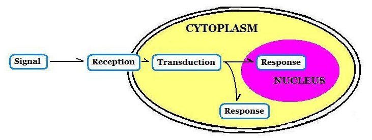
: cells need to communicate with each other to defend against pathogens and exterior environment and to grow and develop.
1. Signal
a. Direct signal and connection
Cell types | Classification | Features |
Animal cell | Tight junction | Tightly associated membranes between two cells |
Gap junction | Channels that directly connect the cytoplasm of adjacent cells | |
Desmosomes | Anchoring junctions held together by adhesion proteins | |
Plant cell | Plasmodesmata | Perforated channels in plant cell wall |
b. Local signal
- Paracrine signal
– diffusing signal through extracellular matrix to near target cells.
– local distance
– eg/ Growth factors, clotting factors, histamines, nitric oxide
- Synaptic signal
– releasing neurotransmitter into synapse
c. Long distance signal
- Endocrine signal
– releasing of extracellular signal known as hormones into the bloodstream.
– long distance
– eg/ Estradiol, testosterone, adrenaline, insulin
2. Reception
a. Cell surface reception
: receptor that binds signal on plasma membrane.
- Ion channel linked receptors
– Ligand binding opens or closes an ion channel, changing ion flow across the membrane.
- G-protein linked receptors
– A ligand-bound GPCR activates a heterotrimeric G protein by promoting GDP–GTP exchange on its α subunit.
- Receptor Tyrosine kinases
– Ligand binding activates receptor tyrosine kinases, which use ATP to phosphorylate tyrosine residues and initiate downstream signaling.
b. Intracellular reception
: receptor that binds hydrophobic hormones (steroid hormones) on the inside of a cell.
3. Transduction
a. Phosphorylation cascade
: a sequence of protein phosphorylation events that relays a signal.
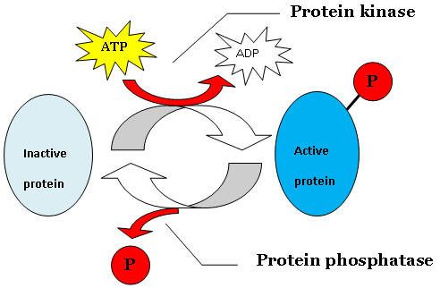
- Phosphorylation
– Protein kinases transfer phosphate groups, usually from ATP, to target proteins.
– Protein phosphatases remove phosphate groups from target proteins.
Phosphorylation can activate or inhibit a protein, depending on the target and site. The figure below illustrates an example in which phosphorylation activates the protein.
b. G-protein signaling pathway
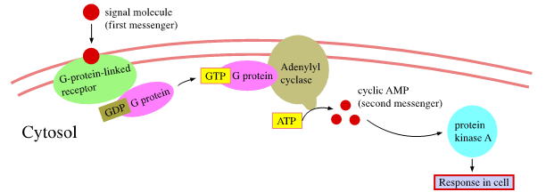
- First messenger (signal molecules): activates G-protein-linked receptor.
- Activated G protein → adenylyl cyclase → cAMP production → protein kinase A activation.
Adenylyl cyclase converts ATP to cAMP, the second messenger.
c. Calcium and IP3 in signaling pathway
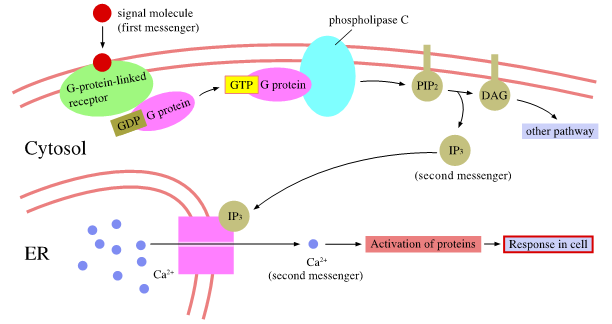
- First messenger (signal molecules): activates G-protein-linked receptor.
- Second messenger (IP3 and Ca2+).
– IP3 binds to IP3-gated calcium channels in the ER membrane, releasing Ca2+ into the cytosol.
– Ca2+ activates various proteins.
4. Response
: leading transcription or cytoplasmic activities
D. Membrane
1. Function
- Separation between inside and outside of cell.
- Selectively regulates transport into and out of the cell. Small hydrophobic molecules can diffuse through the lipid bilayer; ions and many polar solutes require transport proteins.
- Growth and movement.
- Communication with other cells.
2. Components of structure
- Phospholipids – amphipathic molecules with hydrophilic heads and hydrophobic tails, arranged in a bilayer.
- Proteins – integral proteins have membrane-embedded hydrophobic regions; peripheral proteins associate with membrane surfaces.
- Cholesterol – embedded in animal cell membranes and helps regulate membrane fluidity.
- Glycolipids.
3. Passive transport
: net movement of substances down their concentration or electrochemical gradients.
- Does not require direct cellular energy input.
- Examples: simple diffusion, osmosis, and facilitated diffusion through ion channels or carrier proteins.
Chemiosmosis couples proton movement down an electrochemical gradient through ATP synthase to ATP production.
a. Diffusion
- Random molecular motion produces net diffusion from higher to lower concentration.
- Eg/ Movement of O2 and CO2 through cell membrane and movement of solutes substances dissolved in a solution.
b. Osmosis
- Diffusion of water from high water potential area to low water potential area across a semi-permeable membrane.
- Eg/ Water movement through aquaporins of vacuoles.
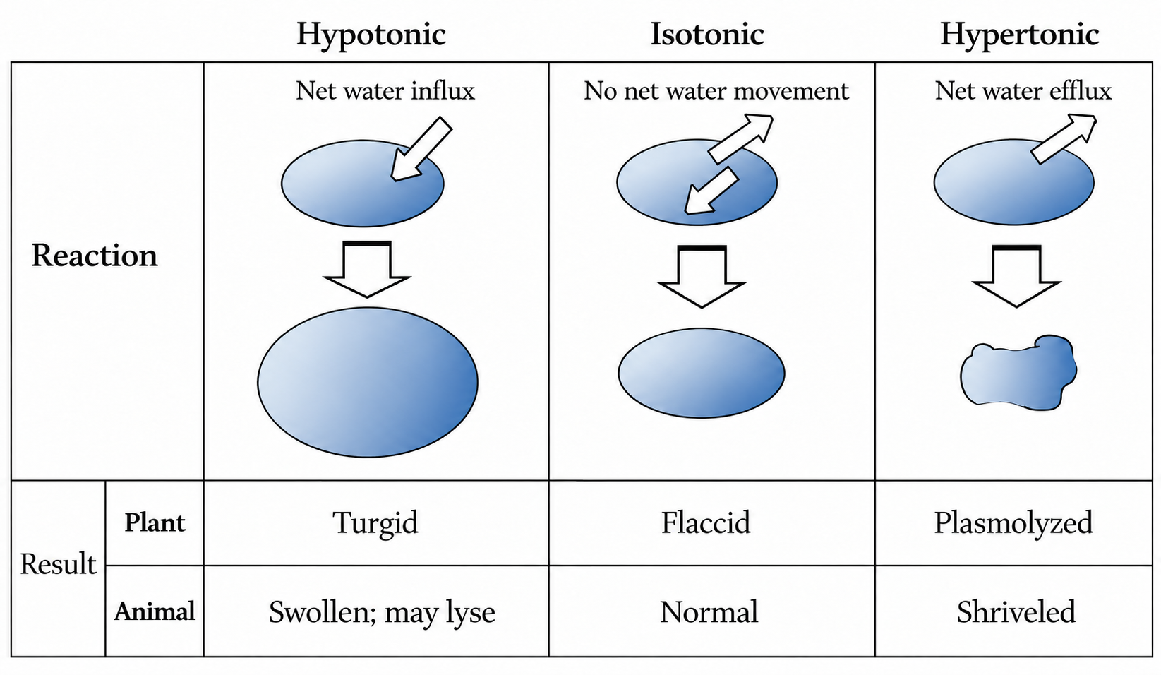
- If cell is placed in
– hypotonic solution → Animal cell: swelling or lysis; plant cell: turgid
* Because of absorption of water, the cell size becomes bigger.
* Eg/ A red blood cell swells up and bursts when it is placed in distilled water.
– isotonic solution → Animal cell: normal; plant cell: flaccid
– hypertonic solution → Animal cell: shriveled; plant cell: plasmolyzed
* Because of losing water, the cell size shrinks.
4. Active transport
: Active transport moves substances against their concentration or electrochemical gradients using energy, either directly from ATP or indirectly from an ion gradient.
- Primary active transport uses energy directly; secondary active transport uses energy stored in an ion gradient.
- Eg/ Sodium-potassium pump of axon, electrogenic pump, cotransport (symport and antiport), proton pump.
5. Bulk transport
: movement of large molecules across the membrane.
Classification | Features | |
Endocytosis | Pinocytosis | Engulfing liquid by using vesicle of membrane |
Phagocytosis | Engulfing particles by using of membrane | |
Receptor-mediated endocytosis | Engulfing molecules by specific receptor of vesicle membrane | |
Exocytosis | Ejection of stuff in vesicle by plasma membrane | |
E. Energy and life
1. Metabolism
: Metabolism is the sum of all chemical reactions in a living organism, including catabolic and anabolic pathways.
- Catabolic pathways – breakdown pathway or digestive process.
- Anabolic pathways – the process of building complicated molecules from smaller units by using energy.
2. Laws of energy
a. First law of thermodynamics
- Principle of conservation of energy – energy can be transferred and transformed, but not created or destroyed.
- Eg/Chemical energy converts into kinetic energy after eating food.
b. Second law of thermodynamics
- Universal law of increasing entropy.
- Eg/ Heat energy emit when organisms work.
3. Homeostasis
: control system to maintain the stable internal condition from outside environmental changes.
- Negative feedback.
– responses oppose deviations from a set point.
– most homeostatic control systems use negative feedback.
- Positive feedback – responses amplify the initial change.
F. Enzymes
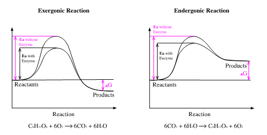
: biological catalysts (mostly proteins) that speed chemical reactions by lowering activation energy without being consumed.
- Substrate binding can induce a conformational change in the enzyme.
- Enzymes speed the approach to equilibrium without changing Keq or the reaction’s ΔG.
- Enzyme activity depends on temperature, pH, and enzyme and substrate concentrations.
- Each enzyme has an optimum temperature and pH.
- Enzymes can be reused after catalysis.
1. Reaction
- Exergonic reaction: release of free energy.
- Endergonic reaction: obtaining free energy.
2. Structure of enzyme
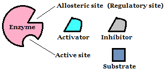
- Allosteric site
– meaning different site.
– regulatory molecules (activator, inhibitor) binding site.
- Active site – substrate binding site
3. Regulation of enzyme
a. Allosteric Enzyme
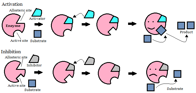
: enzyme can be active or inactive by the binding of regulatory molecule to allosteric site.
- Activation – binding of an activator at an allosteric site increases enzyme activity.
- Inhibition – binding of an inhibitor at an allosteric site reduces enzyme activity.
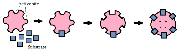
- Positive cooperativity.
– binding of substrate to one subunit increases substrate affinity at other subunits.
– amplify the activity of enzyme.
b. Enzyme with inhibition
- Non-competitive inhibition
– A noncompetitive inhibitor reduces enzyme activity by binding at a site distinct from the active site; it does not necessarily prevent substrate binding.
- Competitive inhibition
– A competitive inhibitor binds to the active site and competes with the substrate.
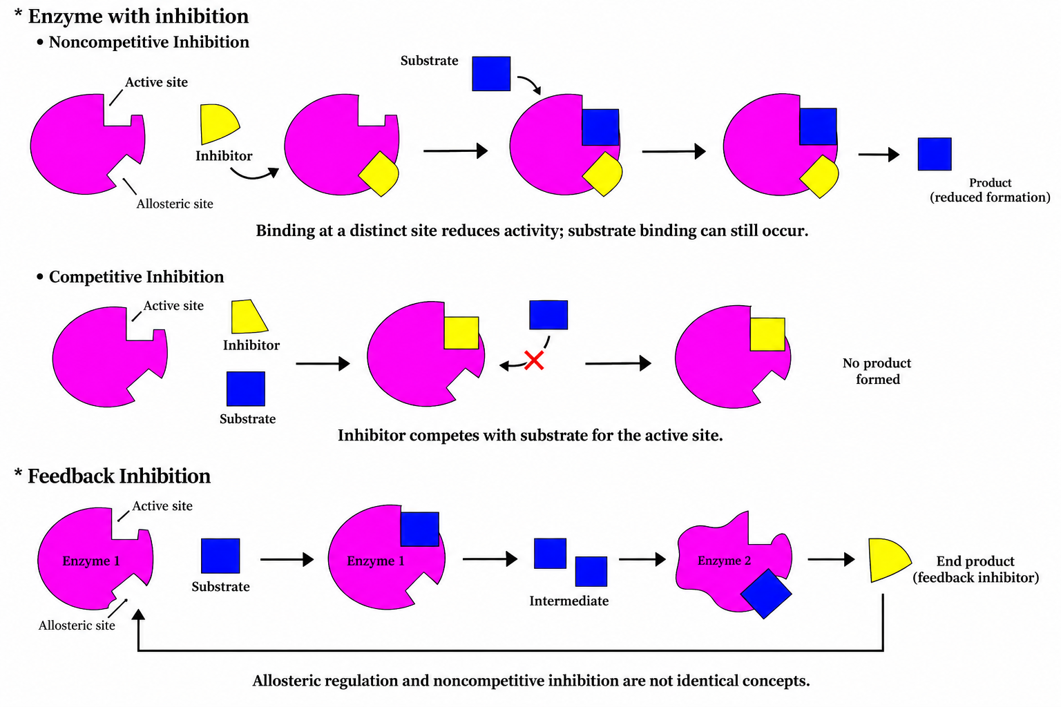
Allosteric regulation includes activation or inhibition at regulatory sites; it is not identical to noncompetitive inhibition.
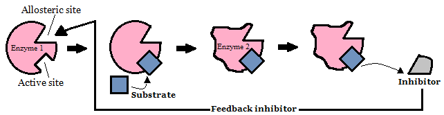
Feedback inhibition
– the end product of a long series of enzymatic reaction inhibits the beginning reaction of the series.
G. Photosynthesis
1. Redox reaction
a. Oxidation
- Losing electron and hydrogen.
- Gaining oxygen.
b. Reduction
- Gaining electron and hydrogen.
- Losing oxygen.
** OILRIG – Oxidation Is Losing electron and Reduction Is Gaining electron.
2. Chloroplast
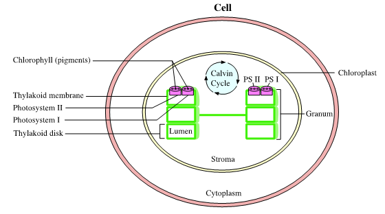
: double membraned organelle of cell, which is to convert light energy to chemical energy.
a. Structure of chloroplast
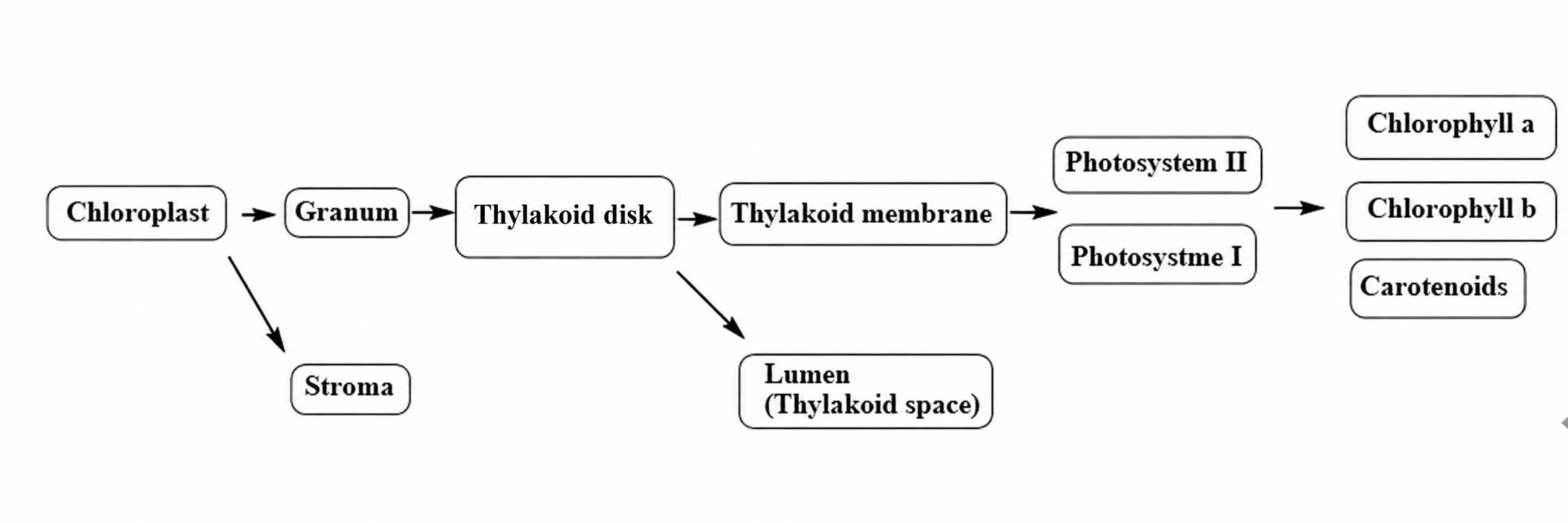
b. Larger to smaller structure
c. Photosynthesis
: converting light energy to ATP free energy and reducing NADP to NADPH to make sugar as a final product.
- Light + 6H2O + 6CO2 → C6H12O6 + 6O2
- Occurring in some bacteria, algae, and plants.
i. Light dependent (light reaction)
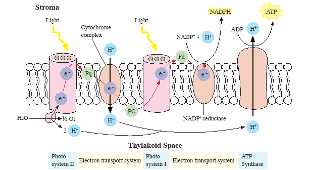
- Taking place at thylakoid membrane.
- Photosynthetic pigments (light receptors).
: magnesium-containing porphyrin ring and hydrocarbon tail in Chlorophyll.
– chlorophyll a: primary pigment that absorbs primarily blue-violet and red light and reflects relatively more green light.
– chlorophyll b: accessory pigment that broadens the range of light absorbed for photosynthesis.
– carotenoid: accessory pigment that absorbs mainly blue-green wavelengths and contributes to photoprotection (protection of chlorophyll by dissipating excessive light energy).
- H2O is source of electron, hydrogen, and oxygen at photosystem II.
2H2O → O2 + 4H+ + 4e−
- PSII oxidizes water and supplies electrons to the photosynthetic electron transport chain, contributing to proton-gradient formation.
- PSI transfers excited electrons to ferredoxin, enabling NADP+ reduction to NADPH.
- Electron transport through the cytochrome complex drives proton movement from the stroma into the thylakoid lumen.
- ATP synthase uses the proton gradient across the thylakoid membrane to synthesize ATP as H+ flows into the stroma.
ii. Calvin cycle (light-independent reactions)
- Takes place in stroma.
- Uses ATP and NADPH to produce sugar in Calvin cycle.
- Calvin cycle
RuBP (Ribulose bisphosphate) + CO2 (by Rubisco enzyme)
→ Two 3-phosphoglycerate (PGA) per turn of Calvin cycle
- Glyceraldehyde 3-phosphate (G3P, also called PGAL) is the net carbohydrate product. Three CO2 fixed yield one net G3P; six yield two net G3P for carbohydrate synthesis. Light-independent does not mean that these reactions occur only in darkness.

d. Photosynthesis in C3, C4, CAM plants
- C3 plants
– Eg/ Most crops (rice, wheat, soybeans).
- C4 plants
– adaptation in hot and dry conditions.
– spatial separation: CO2 fixation in mesophyll cell and then working of Calvin cycle in bundle sheath cell.
– eg/ sugarcane, corn.
- CAM plants
– adaptation in arid conditions.
– temporal separation (night, day) – CO2 enter into stomata during night and then working of Calvin cycle during day.
– open stomata during the night and close stomata during the day.
– eg/ Cacti, pineapples.
H. Cellular respiration
: metabolic reaction, which is to convert nutrients into energy in organism cells
- Aerobic respiration: glycolysis, pyruvate oxidation, the citric acid cycle, and oxidative phosphorylation. Fermentation provides an alternative means of regenerating NAD+ for glycolysis.
C6H12O6 + 6O2 → 6H2O + 6CO2 + Energy (ATP)

1. Glycolysis
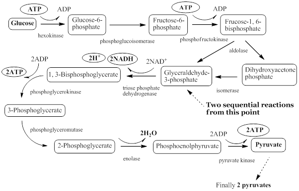
: metabolic pathway, which is to convert one glucose into two pyruvates in cytosol.
- Investing 2ATP and harvesting 4ATP, therefore harvesting net 2ATP.
- Harvesting 2NADH, 2H+, 2H2O, 2Pyruvates.
Glucose + 2NAD+ + 2Pi + 2ADP → 2Pyruvates + 2NADH + 2ATP(NET) + 2H+ + 2H2O
2. Fermentation
: Fermentation regenerates NAD+ from NADH, allowing glycolysis to continue without an electron transport chain.
ATP is produced by substrate-level phosphorylation in glycolysis, not by the fermentation reactions themselves. Fermentation differs from anaerobic respiration, which uses an electron transport chain.

- Lactic acid fermentation converts pyruvate to lactate; alcoholic fermentation produces ethanol and CO2.
- Lactate fermentation occurs in muscle cells; alcoholic fermentation occurs in yeast.
- In the Cori cycle, lactate travels to the liver and is used to regenerate glucose.
- Both fermentation pathways regenerate NAD+.
3. Mitochondria
: double membraned organelle of cell, which is place for oxidative metabolism to make ATP.
- Aerobic respiration
a. Structure of mitochondria
- Double membrane
- Cristae
- Matrix
- Krebs cycle
b. Pyruvate oxidation (acetyl-CoA formation)
- A separate step before the Krebs cycle, occurring in the mitochondrial matrix in eukaryotic cells.
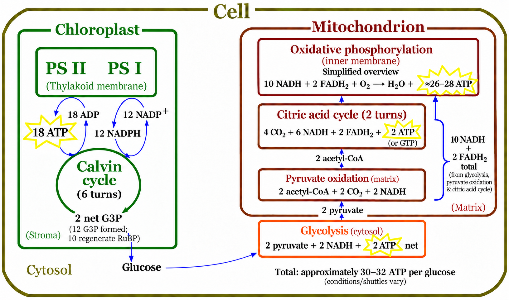
- Per glucose: 2 pyruvate → 2 acetyl-CoA + 2 CO2 + 2 NADH.
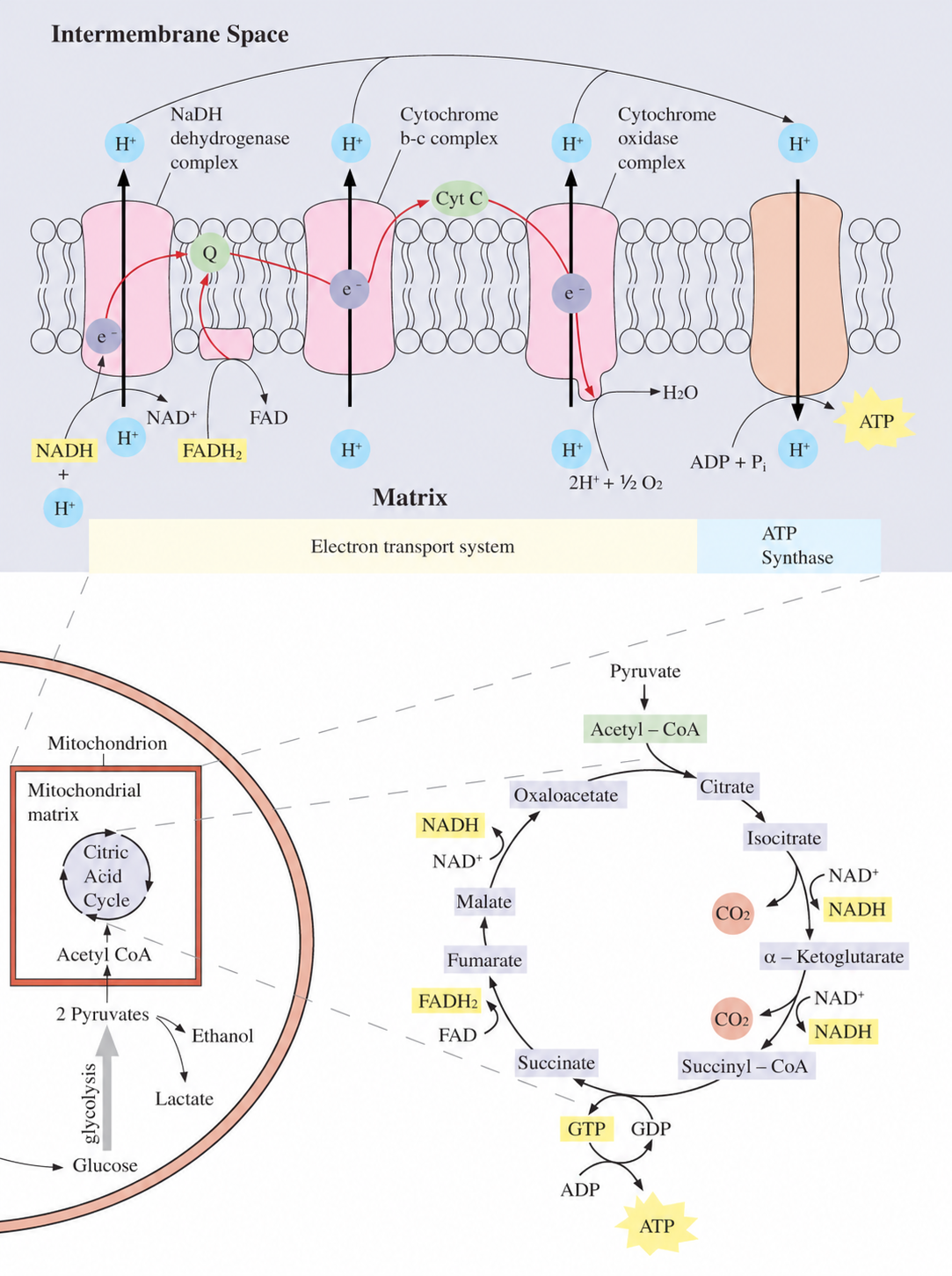
c. Krebs cycle (Citric acid cycle, TCA cycle)

- Aerobic metabolism in mitochondria
- Acetyl CoA + oxaloacetate → citrate
- Producing 6 NADH, 2 FADH2, 2 ATP (2 GTP), and 4 CO2 per 2 acetyl-CoA (derived from one glucose); pyruvate oxidation separately produces 2 NADH and 2 CO2
d. Catabolism of various macromolecules
- Gluconeogenesis – glucose generation of metabolic pathway from amino acid, glycerol, and lactate.

e. Electron transport
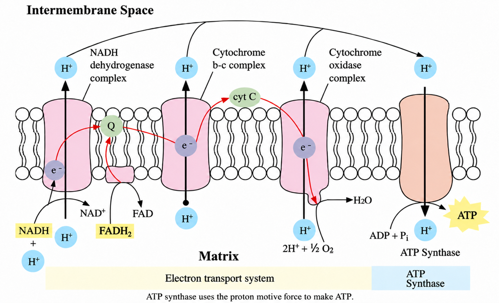
- Electron transport chain and chemiosmosis at inner mitochondrial membrane are called oxidative phosphorylation.
- Ubiquinone (coenzyme Q) is a lipid-soluble mobile electron carrier within the inner mitochondrial membrane.
- Cytochrome c is a small, water-soluble peripheral protein that transfers electrons between complexes III and IV.
- Electron transport chain pumps protons from matrix to intermembrane space to increase a proton gradient.
- ATP synthase uses the proton motive force to synthesize ATP.
- Oxidative phosphorylation typically produces approximately 26–28 ATP per glucose in eukaryotic cells.
- Typical P/O ratios: NADH ≈ 2.5 ATP; FADH2 ≈ 1.5 ATP.
- O2 is the final electron acceptor and is reduced to H2O.

4. Overall reaction between photosynthesis and cellular metabolism
a. Chloroplasts
- Light reactions: PSII releases O2; electron transport and ATP synthase generate ATP, while PSI supports NADPH production.
- For 6 CO2 fixed, the Calvin cycle consumes 18 ATP and 12 NADPH.
- The cycle forms 12 G3P (PGAL); 10 regenerate RuBP and 2 are net products available for carbohydrate synthesis.
b. Mitochondria
- Krebs cycle: 4 CO2, 6 NADH, 2 ATP (or GTP), and 2 FADH2 per 2 acetyl-CoA (one glucose)
- Oxidative phosphorylation: approximately 26–28 ATP per glucose; O2 is reduced to H2O
c. ATP yield from aerobic respiration
- Glycolysis: 2 ATP
- Krebs cycle: 2 ATP
- Oxidative phosphorylation: approximately 26–28 ATP
Total ATP yield from aerobic respiration is approximately 30–32 ATP per glucose, depending on the shuttle system and cellular conditions.
d. Mitochondria and chloroplasts
- Semiautonomous containing DNA and ribosome
- Endosymbiosis